

 PDF
PDF ePub
ePub Citation
 Print
Print


In contrast to the intervertebral disc calcification in adults, pediatric intervertebral disc calcification is a rare disorder. It has been reported less than 200 cases in the literature worldwide and no single case in Korea. It is characterized by calcification of the nucleus pulposus of intervertebral discs. Some cases are asymptomatic and found on incidental radiographs but others are associated with distinct clinical symptoms.1) The etiology of disc calcification is still unclear but it is known to have self-limiting course and excellent prognosis. We present two cases of symptomatic intervertebral disc calcification of cervical spine in children, including the results of laboratory tests, changes of clinical symptoms and radiological findings until spontaneous resolution.
CASE REPORTS
Case 1
A 10-year-old girl visited out-patient clinic with the chief complaint of neck pain. The neck pain started five days ago without any history of preceding trauma or infection. The neck pain was aggravated for five days. She was previously healthy without any skeletal deformities. On the physical examination, pain and tenderness were localized on the C2-3 and C5-6 intervertebral discs. Neck motion was mildly restricted due to pain without torticollis. She did not complained radiating pain and free of neurologic deficit.
Laboratory findings revealed a total white blood cell count of 9,920/mm3, an erythrocyte sedimentation rate of 2 mm/hr, a C-reactive protein of 0.04 mg/dl, which were in normal range without any evidence of infection. Serum calcium was 8.9 mg/dl, phosphorus, 5.1 mg/dl and alkaline phosphatase, 240 IU/L. Kidney and liver function tests were also within normal limits. Any metabolic bone diseases and conditions prone to heterotopic bone formations could be ruled out by lab findings above.
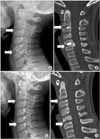
Initial plain radiograph showed densely calcified discs in C2-3 and C5-6 (Fig. 1A). Calcified disc of C2-3 was smaller and round in shape; however, that of C5-6 was larger, fragmented and herniated (Fig. 1B). The patient was treated with steroid (Tracinon® 1 mg) and non-steroidal anti-inflammatory drug (Rhonal® 100 mg) initially for a week and became asymptomatic five days later. Follow-up radiograph and CT scan one and a half year later revealed that calcified discs of the C2-3 and C5-6 had completely resolved (Fig. 1C, D).
Case 2
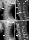
A 12-year-old boy presented with torticollis without any neurologic symptoms or signs that had begun two weeks ago. No preceding trauma history or infection was noted. On examination, the head was tilted to the right and changes of neck position caused severe neck pain. Laboratory findings revealed a total white blood cell count of 5,360/mm3, an erythrocyte sedimentation rate of 20 mm/hr and a C-reactive protein of 0.08 mg/dl. Serum levels of calcium, phosphorus and alkaline phosphatase were also within normal limits. Initial plain radiograph and MRI showed densely calcified disc of the C5-6 which was confined to nucleus pulposus (Fig. 2A, B). He was treated with non-steroidal anti-inflammatory drug (Rhonal® 100 mg) for 10 days and cervical collar. Torticollis and neck pain were completely resolved in one month. Plain radiograph and CT scan taken one year later revealed that calcified disc of the C5-6 markedly decreased (Fig. 2C, D).
DISCUSSION
The etiology of cervical disc calcification in children has been postulated to be trauma,2) infection or inflammation,3) but is still obscure. Smith et al3) performed an anterior discectomy in a 12-year old boy at the C4-5 and demonstrated an inflammatory response of severe reactive fibroblastic proliferation. Contrary to this, Swick4) found no evidence of inflammatory or reactive changes of neovascularization. Trauma has often been implicated as a predisposing factor because disc calcification in some children was preceded by trauma.2) However, preexisting disc calcification was also identified in some children without history of trauma. We studied 2 cases to rule out any conditions known to cause hetrotopic calcification, which was negative. Thus, we could not find any relationship between trauma, infection or metabolic bony disorder in our cases with cervical disc calcification in children.
The clinical manifestations of disc calcification are variable. By far the most common symptom is neck pain without a history of trauma as seen in our cases.5) There is no definite explanation for the cause of neck pain. Bagatur et al6) suggested that the clinical symptoms were related to an inflammatory process within the disc. Another hypothesis suggests that neck pain is associated with a rise in intradiscal pressure.7) Torticollis is another fairly common symptom, occurring in 40% of symptomatic patients as in our second case.
Neurologic deficits such as radicular pain, focal weakness and myopathy5) occur very infrequently. However, herniation of calcified discs occasionally leads to acute nerve root or spinal cord compression.5) Sonnabend et al7) stated that the incidence of disc herniation was up to 38% of patients with symptomatic intervertebral disc calcification, but the actual neurologic symptoms were rare in these patient group. Thus, It is assumed that herniation of calcified discs are not always correlated with neurologic symptoms.
Single cervical level was involved in eighty five to ninety percent of patients.6) One or two levels were involved in our case. Dai et al2) suggested that plain radiographs were sufficient to assess disc calcification in children, and additional investigation with imaging may be recommended only for those with a neurological deficit. But, the plain radiographs cannot always identify the extent and herniation of the calcified disc as in our first case (Fig. 1B).
All the previous authors2,4,6) reported that the symptoms were resolved earlier than the radiographic resolution of calcification. Dai et al2) reported that the mean time to resolution of the symptoms was 34.3 days (range, seventeen to 173 days) and the mean time to resolution of calcification was fifteen months (range, three to sixty months). In our first case, cervical disc calcification was completely resolved in one year six months. In second case, disc calcification markedly decreased at one year follow up CT scan. Thus, radiologic improvement takes longer time than clinical improvement, even several months to years in our cases.
The natural history of cervical disc calcification in children is excellent and surgical treatment is rarely indicated.2,8) Most of children respond well to the conservative treatments within several months including analgesics and cervical orthosis as in our cases. Surgical intervention has been reported only for few cases with a neurologic deficit.9) The indications of surgical decompression might be intractable neck pain with progression of neurologic deficits.
Cervical intervertebral disc calcification in children is a rare disorder with good prognosis. It must be included for the differential diagnosis for the neck pain or torticollis in children. Any physicians or surgeons should understand the natural history for treating these children.
 XML Download