

 PDF
PDF ePub
ePub Citation
 Print
Print


Abstract
Purpose
We wanted to evaluate the clinical and radiological results of one-staged open-wedge high tibial osteotomy (HTO) and arthroscopic anterior cruciate ligament (ACL) reconstruction for patients with complete rupture of the ACL and concomitant varus malalignment of the lower limb.
Materials and Methods
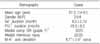
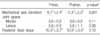
Twenty-five patients were prospectively assessed before and 1 year after their simultaneous operation as a single procedure. The clinical assessment included the Lysholm score, the Tegner activity level scale and a physical examination. The radiological data was used to calculate the mechanical axis, the joint space, the tibial slope and the arthrometric stress test. The postoperative complications were also assessed.
Results
The Lysholm knee score and the Tegner activity level scale improved from a mean of 72.1 to 93.8 and from 1.45 to 4.65, respectively (p<0.05). According to the Lachmann test and the pivot shift test for stability, improvements were made to a grade of 0 or I in most of the patients (p<0.05) and the STSD with using an arthrometric device significantly improved from 8.8 mm to postoperative 2.6 mm (p<0.05). The mechanical axis significantly improved from varus 6.1 degrees to valgus 0.3 degrees (p<0.05). Osteoarthritis of the knee, the posterior tibial slope and the joint space did not show any significant changes (p>0.05). The severity and rate of the postoperative complications were both low.
Conclusion
One-staged open-wedge HTO and ACL reconstruction produced satisfactory correction of the mechanical axis alignment and, it improved knee function. Further, the procedure had a low complication rate. It is also cost effective due to, reducing the frequency of operation and avoiding overlap of rehabilitation.
Figures and Tables
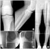 | Fig. 2This radiographs show the preoperative antero-posterior and lateral radiography (A), the teleoroentgenography (B) and the STSD with using Telos® device (C); the posterior slope was 8.5°, the mechanical axis deviation was varus 7°, and the STSD was 7 mm at 20 Lbs.
|
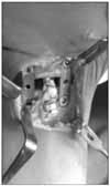 | Fig. 3This intra-operative photograph shows the ACL graft after fixation in tibial tunnel with bio-interference screw with additional staple after open-wedge HTO.
|
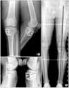 | Fig. 4This radiographs show the postoperative antero-posterior and lateral radiography (A), and the teleoroentgenography (B) and the STSD after 1 year post-operation (C); the posterior slope was 9°, the mechanical axis deviation was varus 0°, and the STSD was 3 mm at 20 Lbs. (A): lateral cortex without breakage is preserved and height of anterior plate is 70% compared to posterior plate to prevent increase of the posterior slope. In addition, well placed Endo-button is shown.
|


References
1. Noyes FR, Barber SD, Simon R. High tibial osteotomy and ligament reconstruction in varus angulated, anterior cruciate ligament-deficient knees. A two- to seven-year follow-up study. Am J Sports Med. 1993. 21:2–12.
2. Giffin JR, Shannon FJ. The role of the high tibial osteotomy in the unstable knee. Sports Med Arthrosc. 2007. 15:23–31.
3. Badhe NP, Forster IW. High tibial osteotomy in knee instability: the rationale of treatment and early results. Knee Surg Sports Traumatol Arthrosc. 2002. 10:38–43.

4. van de Pol GJ, Arnold MP, Verdonschot N, van Kampen A. Varus alignment leads to increased forces in the anterior cruciate ligament. Am J Sports Med. 2009. 37:481–487.
5. Holden DL, James SL, Larson RL, Slocum DB. Proximal tibial osteotomy in patients who are fifty years old or less. A long-term follow-up study. J Bone Joint Surg Am. 1988. 70:977–982.
6. Dejour H, Neyret P, Boileau P, Donell ST. Anterior cruciate reconstruction combined with valgus tibial osteotomy. Clin Orthop Relat Res. 1994. 299:220–228.
7. Lattermann C, Jakob RP. High tibial osteotomy alone or combined with ligament reconstruction in anterior cruciate ligament-deficient knees. Knee Surg Sports Traumatol Arthrosc. 1996. 4:32–38.
8. Noyes FR, Barber-Westin SD, Hewett TE. High tibial osteotomy and ligament reconstruction for varus angulated anterior cruciate ligament-deficient knees. Am J Sports Med. 2000. 28:282–296.
9. Song EK, Seon JK, Park SJ. How to avoid unintended increase of posterior slope in navigation assisted open-wedge high tibial osteotomy. Orthopedics. 2007. 30:Suppl. 127–131.
10. Fried JA, Bergfield JA, Weiker G, Andrish JT. Anterior cruciate reconstruction using the Jones-Ellison procedure. J Bone Joint Surg Am. 1985. 67:1029–1033.
11. Bartlett RJ, Crowe R. Results of intra-articular anterior cruciate ligament reconstruction using patellar ligament. J Bone Joint Surg Br. 1984. 66:788.
12. Insall JN, Joseph DM, Msika C. High tibial osteotomy for varus gonarthrosis. A long-term follow-up study. J Bone Joint Surg Am. 1984. 66:1040–1048.
13. Noyes FR, Schipplein OD, Andriacchi TP, Saddemi SR, Weise M. The anterior cruciate ligament-deficient knee with varus alignment. An analysis of gait adaptations and dynamic joint loadings. Am J Sports Med. 1992. 20:707–716.
14. Marti RK, Verhagen RA, Kerkhoffs GM, Moojen TM. Proximal tibial varus osteotomy. Indications, technique, and five to twenty-one-year results. J Bone Joint Surg Am. 2001. 83:164–170.
15. Giffin JR, Vogrin TM, Zantop T, Woo SL, Harner CD. Effects of increasing tibial slope on the biomechanics of the knee. Am J Sports Med. 2004. 32:376–382.
16. Hohmann E, Bryant A, Imhoff AB. The effect of closed wedge high tibial osteotomy on tibial slope: a radiographic study. Knee Surg Sports Traumatol Arthrosc. 2006. 14:454–459.
17. Marti CB, Gautier E, Wachtl SW, Jakob RP. Accuracy of frontal and sagittal plane correction in open-wedge high tibial osteotomy. Arthroscopy. 2004. 20:366–372.
18. Lobenhoffer P, Agneskirchner JD. Improvements in surgical technique of valgus high tibial osteotomy. Knee Surg Sports Traumatol Arthrosc. 2003. 11:132–138.
19. Agneskirchner JD, Hurschler C, Stukenborg-Colsman C, Imhoff AB, Lobenhoffer P. Effect of high tibial flexion osteotomy on cartilage pressure and joint kinematics: a biomechanical study in human cadaveric knees. Winner of the AGA-Donjoy Award 2004. Arch Orthop Trauma Surg. 2004. 124:575–584.
 XML Download