

 PDF
PDF ePub
ePub Citation
 Print
Print


Abstract
Purpose
We wanted to evaluate the mechanical strength of proximal tibia as resection distance increased from the joint surface.
Materials and Methods
We obtained the CT images of twenty knee osteoarthritis patients undergoing total knee arthroplasty. The finite element models were created based on the computed tomography images. The 8-node hexahedron element was made from BIONIX™ (CANTIBio. Co, Suwon, Korea), which is automatic mesh generation software program. The finite element model of the proximal tibia was resected at 6 mm, 8 mm, 10 mm, 12 mm, 15 mm and 18 mm from the lateral joint surface. A 1% strain rate was applied to a model by using HyperMesh™ software (Altair Engineering. Inc, Seattle, USA). The ultimate stress was calculated from the finite element analysis with using ANSYS 9.0 (ANSYS. Inc, Orlando, USA).
Results
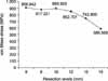
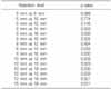
The mean ultimate stress was 906.84 MPa, 877.22 MPa, 895.93 Mpa, 852.70 MPa, 742.90 Mpa and 585.51 Mpa at the 6 mm, 8 mm, 10 mm, 12 mm, 15 mm and 18 mm resection levels. As compare to the 6 mm resection level, the bone strengths at 15 mm and 18 mm were decreased with statistical significance (15 mm: p=0.005, 18 mm: p=0.000).
Figures and Tables
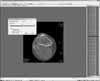
Fig. 2
Volumetric mesh model from computed tomographic data. (A) Results of region-growing algorithm. (B) Voxel size: X: 0.163 mm, Y: 0.163 mm, Z: 1.000 mm.
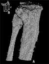
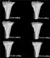
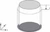
Fig. 4
Boundary condition. A strain was applied at the top face (◯) while displacement was constrained at the bottom face (◯).
References
1. Mckinley TO, Bay BK. Trabecular bone strain changes associated with subchondral stiffening of the proximal tibia. J Biomech. 2003. 36:155–163.

2. Chaput CD, Weeden SH, Hyman WA, Hitt KD. Mechanical bone strength of the tibial resection surface at increasing distance from the joint line in total knee arthroplasty. J Surg Orthop Adv. 2004. 13:195–198.
3. Goldstein SA, Wilson DL, Sonstegard DA, Matthews LS. The mechanical properties of human tibial trabecular bone as a function of metaphyseal location. J Biomech. 1983. 16:965–969.
4. Harada Y, Wevers HW, Cooke TD. Distribution of bone strength in the proximal tibia. J Arthroplasty. 1988. 3:167–175.
5. Hvid I. Mechanical strength of trabecular bone at the knee. Dan Med Bull. 1988. 35:345–365.
6. Hvid I, Jensen J, Nielsen S. Bone strength measurements at the proximal tibia. Penetration tests and epiphyseal compressive strength. Int Orthop. 1986. 10:271–275.
7. van Rietbergen B, Weinans H, Huiskes R, Polman BJW. Computational strategies for iterative solutions of large FEM applications employing voxel data. Int J Num Meth Eng. 1998. 39:2743–2767.
8. Odgaard A, Linde F. The underestimation of Young's modulus in compressive testing of cancellous bone specimens. J Biomech. 1991. 24:691–698.
9. van Rietbergen B, Ulrich D, Pistoia W, Huiskes R, Ruegsegger P. Trabecular bone ultimate stress can be predicted from large-scale FE-analyses. J Biomech. 1998. 31:Suppl 1. 151.
10. van Rietbergen B, Weinans H, Huiskes R, Odgaard A. A new method to determine trabecular bone elastic properties and loading using micromechanical finite-element models. J Biomech. 1995. 28:69–81.
11. Ritter MA, Montgomery TJ, Zhou H, Keating ME, Faris PM, Meding JB. The clinical significance of proximal tibial resection level in total knee arthroplasty. Clin Orthop Relat Res. 1999. 360:174–181.
12. Bentzen SM, Hvid I, Jørgensen J. Mechanical strength of tibial trabecular bone evaluated by X-ray computed tomography. J Biomech. 1987. 20:743–752.
13. Bourne BC, van der Meulen MC. Finite element models predict cancellous apparent modulus when tissue modulus is scaled from specimen CT-attenuation. J Biomech. 2004. 37:613–621.
14. Sarathi Kopparti P, Lewis G. Influence of three variables on the stresses in a three-dimensional model of a proximal tibia-total knee implant construct. Biomed Mater Eng. 2007. 17:19–28.
15. Majumdar S, Kothari M, Augat P, et al. High-resolution magnetic resonance imaging: Three-dimensional trabecular bone architecture and biomechanical properties. Bone. 1998. 22:445–454.
 XML Download