

 PDF
PDF ePub
ePub Citation
 Print
Print


Abstract
Purpose
To report the long-term follow-up results of a free vascularized fibular graft (FVFG) for the treatment of patients with congenital pseudarthrosis of the tibia (CPT).
Materials and Methods
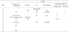
Fourteen patients with CPT, who underwent FVFG and were followed-up for more than 4 years, were enrolled in this study. The average age at FVFG was 4.5 years (range, 1~10.6 years) with an average follow-up of 11.6 years (range, 4~26.6 years). In 11 cases, 24 additional procedures were performed due to the secondary problems, such as delayed union, nonunion, leg length discrepancy, and deformity of the tibia. The clinical and radiological data were reviewed to evaluate the outcomes of the FVFG.
Results
Bone union and hypertrophy of the fibular graft was observed in all cases. The average time of union in 7 cases, in whom bone union was achieved without the aid of any supplementary procedures, was 6.7 months (range, 3.5~11 months). At the last follow-up, the mean limb length discrepancy was 2.5 cm (range, 0~8 cm), and the average anterior apex and valgus angulation of the tibial shaft were 18.4° (range, 0~85°) and 9.9° (range, -5~34°), respectively. The mean ankle alignment was valgus 7.2° (range, -3~30°), and the mean range of motion of the ankle joint was dorsiflexion 17° and plantar flexion 30°.
Conclusion
FVFG is an effective procedure in congenital pseudarthrosis of the tibia which has the advantages of allowing a more aggressive resection of the abnormal bone and scar tissue and as well as resolving the bone defect after resecting the lesion. This procedure is expected to produce a better clinical result if rigid internal and external fixation are performed.
Figures and Tables
Fig. 1
Case 4. (A) Bony union was failed after bone graft at other hospital. (B) Postoperative radiograph. (C) Seven months after FVFG, solid bony union was obtained. (D) One year nine months after FVFG, distal tibiofibular synostosis was performed due to valgus deformity of the ankle at recipient site. (E) Radiographs of the postoperative 8 year 9 months.
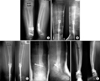
Fig. 2
Case 6. (A) Preoperative radiograph. (B) Postoperative radiograph. (C) Nine months after vascularized fibular graft. (D) Radiographs of the postoperative 8 year 9 months.
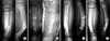
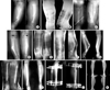
Fig. 3
Case 3. (A) Initial radiograph. (B) At other hospital, bone graft was performed and bony union was failed. (C) Postoperative radiograph. (D) Five months after FVFG, delayed union at proximal part was observed. (E) Two years four months after FVFG, distal tibiofibular synostosis was performed due to valgus deformity of the ankle at donor site. (F) Five years after FVFG, distal tibiofibular synostosis was performed at recipient site. (G) Radiograph prior to corrective osteotomy showing incomplete bony union and deformity of the tibia. Anterior apex and valgus angulation of the tibial shaft were 50 and 20 degree, respectively. (H) Eight years after FVFG, corrective osteotomy and bone graft, intramedullary rod fixation were performed. (I) Scanogram prior to bone lengthening using ilizarov external fixator showed the leg length discrepancy (LLD) about 4.5 cm. (J) Seventeen years after FVFG, bone lengthening was performed. (K) Twenty Five years nine months after FVFG, although LLD was resolved, however the valgus ankle deformity was shown.
References
1. Andersen KS. Congenital pseudarthrosis of the tibia and neurofibromatosis. Acta Orthop Scand. 1976. 47:108–111.

2. Chen CW, Yu ZJ, Wang Y. A new method of treatment of congenital tibial pseudoarthrosis using free vascularized fibular graft: a preliminary report. Ann Acad Med Singapore. 1979. 8:465–473.
3. Dobbs MB, Rich MM, Gordon JE, Szymanski DA, Schoenecker PL. Use of an intramedullary rod for treatment of congenital pseudarthrosis of the tibia. A long-term follow-up study. J Bone Joint Surg Am. 2004. 86:1186–1197.
4. EI-Gammal TA, EI-Saved A, Kotb MM. Telescoping vascularized fibular graft: a new method for treatment of congenital tibial pseudarthrosis with severe shortening. J Pediatr Orthop B. 2004. 13:48–56.
5. Ghanem I, Damsin JP, Carlioz H. Ilizarov technique in the treatment of congenital pseudarthrosis of the tibia. J Pediatr Orthop. 1997. 17:685–690.
6. Gilbert A, Brochman R. Congenital pseudarthrosis of the tibia. Long-term follow-up of 29 cases treated by microvascular bone transfer. Clin Orthop Relat Res. 1995. 314:37–44.
7. Kim HW, Weinstein SL. Intramedullary fixation and bone grafting for congenital pseudarthrosis of the tibia. Clin Orthop Relat Res. 2002. 405:250–257.
8. Minami A, Kato H, Suenaga N, Iwasaki N. Telescoping vascularized fibular graft: a new method. J Reconstr Microsurg. 2003. 19:11–16.
9. Paley D, Catagni M, Argnani F, Prevot J, Bell D, Armstrong P. Treatment of congenital pseudoarthrosis of the tibia using the Ilizarov technique. Clin Orthop Relat Res. 1992. 280:81–93.
10. Peterson D. Congenital pseudarthrosis of the tibia. An overview. Clin Orthop Relat Res. 1989. 247:44–53.
11. Pho RWH. Pho RWH, editor. Free vascularized fibular transplant in management of congenital pseudoarthrosis of the tibia. Microsurgical technique in orthopaedics. 1988. 1st ed. London: Butterworths;152–158.
12. Plawecki S, Carpentier E, Lascombes P, Prevot J, Robb JE. Treatment of congenital pseudarthrosis of the tibia by the Ilizarov method. J Pediatr Orthop. 1990. 10:786–790.
13. Romanus B, Bollini G, Dungl P, et al. Free vascular fibular transfer in congenital pseudoarthrosis of the tibia: results of the EPOS multicenter study. European Paediatric Orthopaedic Society (EPOS). J Pediatr Orthop B. 2000. 9:90–93.
14. Sakamoto A, Yoshida T, Uchida Y, Kojima T, Kubota H, Iwamoto Y. Long-term follow-up on the use of vascularized fibular graft for the treatment of congenital pseudarthrosis of the tibia. J Orthop Surg. 2008. 3:13.
15. Simonis RB, Shirali HR, Mayou B. Free vascularized fibular grafts for congenital pseudarthros is of the tibia. J Bone Joint Surg Br. 1991. 73:211–215.
16. Toh S, Harata S, Tsubo K, Inoue S, Narita S. Combining free vascularized fibula graft and the ilizarov external fixator: recent approaches to congenital pseudarthrosis of the tibia. J Reconstr Microsurg. 2001. 17:497–508.
17. Weiland AJ, Weiss AP, Moore JR, Tolo VT. Vascularized fibular grafts in the treatment of congenital pseudarthrosis of the tibia. J Bone Joint Surg Am. 1990. 72:654–662.
 XML Download