

 PDF
PDF ePub
ePub Citation
 Print
Print


Abstract
Purpose
This study is to evaluate chinical and radiological results of open wedge high tibial osteotomy using Aescula® plate.
Materials and Methods
Ninity one patients who have unicompartmental osteoarthritis with varus deformity were treated by open wedge high tibial osteotomy with Aescula® plate and followed up at least 2 years. Clinically, visual analogue scale (VAS), range of motion (ROM) and hospital for special surgery (HSS) score were evaluated. Radiologically, tibio-femoral angle, mechanical axis, medial proximal tibia angle and posterior slope were measured. All complications were also evaluated.
Results
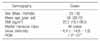
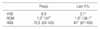
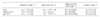
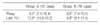
During the follow-up VAS improved from 8.3 to 2.1, ROM were checked preoperatively from 1.3° to 137° and from 1.8° to 136.1° at last follow up. And HSS score improved from 76.8 to 91. Preoperative tibio-femoral angle was 0.4° of varus, mechanical axis 6.4° of varus, medial proximal tibia angle 84.4° and posterior slope 9.3°. Radiologic results at last follow up revealed significant improvements by 8.3° of valgus for tibio-femoral angle, 1.8° of valgus for mechanical axis and 90.1° for medial proximal tibia angle. Mean posterior slope was 10.4° which increased 1.1° compared with preoperative one. And there was one fixation failure that needed re-operation.
Figures and Tables

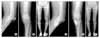 | Fig. 2A comparision of tibial open wedge osteotomy. (A) Preoperative anteroposterior view with tibiofemoral angle. (B) Preoperative lateral view with posterior angle. (C) Teleoroentgenography with mechanical axis. (D) Postoperative antero-posterior view with tibiofemoral angle. (E) Postoperative lateral view with posterior angle. (F) Postoperative teleoroentgenography with mechanical axis.
|
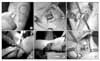 | Fig. 3A procedure of high tibial open wedge osteotomy. (A) Skin incision below medial joint line. (B) Pes anserius tendon is identified and incised with Z-plasty fasion. (C) Identification of anterior border of medial collateral ligament. (D) Guide wire insertion under the fluoroscopy. (E) Osteotomy is performed along guider wire. (F) Gap measeurement and plate fixatioin.
|
References
1. Amendola A, Panarella L. High tibial osteotomy for the treatment of unicompartmental arthritis of the knee. Orthop Clin North Am. 2005. 36:497–504.

2. Berman AT, Bosacco SJ, Kirshner S, Avolio A Jr. Factors influencing long-term results in high tibial osteotomy. Clin Orthop Relat Res. 1991. 272:192–198.
3. Coventry MB. Osteotomy about the knee for degenerative and rheumatoid arthritis. J Bone Joint Surg Am. 1973. 55:23–48.
4. Hernigou P, Medevielle D, Debeyre J, Goutallier D. Proximal tibial osteotomy for osteoarthritis with varus deformity. A ten to thirteen-year follow-up study. J Bone Joint Surg Am. 1987. 69:332–354.
5. Insall JN, Joseph DM, Msika C. High tibial osteotomy for varus gonarthrosis. A long-term follow-up study. J Bone Joint Surg Am. 1984. 66:1040–1048.
6. Katz MM, Hungerford DS, Krackow KA, Lennox DW. Results of total kneearthroplasty after failed proximal tibial osteotomy for osteoarthritis. J Bone Joint Surg Am. 1987. 69:225–233.
7. Koshino T, Murase T, Saito T. Medial opening-wedge high tibial osteotomy with use of porous hydroxyapatite to treat medial compartment osteoarthritis of the knee. J Bone Joint Surg Am. 2003. 85:78–85.
8. Marti RK, Verhagen TA, Kerkhoffs GM, Moojen TM. Proximal tibial varus ostetomy. Indications, technique, and five to twenty-one-year results. J Bone Joint Surg Am. 2001. 83:164–170.
9. Matthews LS, Goldstein SA, Malvitz TA, Katz BP, Kaufer H. Proximal tibial osteotomy. Factors that influence the duration of satisfactory function. Clin Orthop Relat Res. 1988. 229:193–200.
10. Murphy SB. Tibial osteotomy for genu varum. Indications, preoperative planning, and technique. Orthop Clin North Am. 1994. 25:477–482.
11. Niemeyer P, Koestler W, Kaehny C, et al. Two-year results of open-wedge high tibial osteotomy with fixation by medial plate fixator for medial compartment arthritis with varus malalignment of the knee. Arthroscopy. 2008. 24:796–804.
12. Song EK, Seon JK, Park SJ, Seo HY. Navigated open wedge high tibial osteotomy. Sports Med Arthrosc Rev. 2008. 16:84–90.
13. Spahn G. Complication in high tibial (medial opening wedge) osteotomy. Arch Orthop Traum Surg. 2003. 124:649–653.
14. Sundaram NA, Hallett JP, Sullivan MF. Dome osteotomy of the tibia for osteoarthritis of the knee. J Bone Joint Surg Br. 1986. 68:782–786.
15. Van den Bekerom MP, Patt TW, Kleinhout MY, van der Vis HM, Albers GH. Early complications after high tibial osteotomy: a comparison of two techniques. J Knee Surg. 2008. 21:68–74.
16. Windsor RE, Insall JN, Vince KG. Technical considerations of total knee arthroplasty after proximal tibial osteotomy. J Bone Joint Surg Am. 1988. 70:547–555.
 XML Download