Abstract
Purpose
To evaluate the midterm clinical and histological results after autologous chondrocyte implantation (ACI) for an articular cartilage defect of the distal femoral condyle.
Materials and Methods
Twenty four cases with an articular cartilage defect (Outerbridge grade IV) of the femoral condyle that was confirmed by MRI and the arthroscopic findings underwent ACI. Their mean age at the time of surgery was 42.8 years and the mean follow-up period was 53.2 months (range, 20-82 months). At the last follow up, the articular cartilage view (SPGR) of MRI was examined and the clinical results were evaluated using the HSS and Lysholm scores. In 8 cases, second-look arthroscopy and biopsy were performed and evaluated using histological and histochemical methods.
Results
All cases except for one showed well-regenerated articular cartilage on MRI. All cases showed significant clinical improvement in the HSS and Lysholm scores (p<0.0001), with the exception of the Lysholm score of an articular cartilage fracture. Histologically, the regenerated tissue appeared to be a hyaline-like cartilage in all specimens.
Conclusion
ACI for the treatment of articular cartilage defects of the distal femoral condyle showed a good clinical and MRI results. In OA, the clinical results were relatively acceptable after an associated high tibial valgus osteotomy. However, a longer term follow-up study will be needed to reach a final conclusion.
Figures and Tables
 | Fig. 1A gel type cultured chondrocyte was put on the articular cartilage defect of the MFC (A) and was covered with periosteum and sutured to the articular cartilage with 6-0 prolene (B) in the left knee of 59 years old male. |
 | Fig. 2(A) Histogram showing the mean HSS score of OA, OCD and articular cartilage fracture group checked preoperatively and at the last follow-up, respectively. In OA, OCD and articular cartilage fracture group, the HSS scores at the last follow-up were significantly improved than preoperative condition (p<0.0001). (B) Histogram showing the mean Lysholm score of OA, OCD and articular cartilage fracture group checked at preoperative and at the last follow-up, respectively. In OA and OCD, the Lysholm scores at the last follow-up were significantly improved than preoperative condition (p<0.0001). In articular cartilage fracture group, the score was not significantly different between the preoperative and the final follow-up condition (p=0.1257). |
 | Fig. 3A second look arthroscopic biopsy specimen in the left knee of 54 years old female osteoarthritic knee in 3 years and 2 months after operation. Biopsy specimen with 3 mm diameter Turkel needle from the ACI site of medial femoral condyle with close up view of the biopsy site (lower inset). Biopsy specimen at ACI site showed thicker articular cartilage (upper insert A) than normal articular cartilage of the lateral femoral condyle (upper insert B). |
 | Fig. 4Staining of the same specimen shows viable chondrocytes in lacunae with a uniform matrix. (A) The red color of the superficial layer of the biopsy specimen is thinner than the control specimen on safranin-O staining (×40). (B) The Masson's trichrome stain of the biopsy specimen shows a typical distribution of collagen fibers (×40). (C) Immunohistochemical staining for type II collagen shows that the positivity is evenly & diffusely extended to whole layer of the extracellular matrix (×40). |
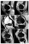
 | Fig. 5MRIs of the MFC of the right OA knee of 59 years old man shows full thickness defect of articular cartilage, preoperatively (A-1) and successful repair (A-2). MRIs of the MFC of the right OCD knee of 20 years old man shows full thickness defect of articular cartilage, preoperatively (B-1) and successful repair at final follow-up (B-2). MRIs of the injured MFC of the right knee of 25 years old man shows full thickness defect of the articular cartilage (C-1) and successful repair at final follow- up (C-2). |
References
1. Binazzi R, Soudry M, Mestriner LA, Insall JN. Knee arthroplasty rating. J Arthroplasty. 1992. 7:145–148.

2. Brittberg M, Lindahl A, Nilsson A, Ohlsson C, Isaksson O, Peterson L. Treatment of deep cartilage defects in the knee with autologous chondrocyte transplantation. N Engl J Med. 1994. 331:889–895.

3. Brittberg M, Winalski CS. Evaluation of cartilage injuries and repair. J Bone Joint Surg Am. 2003. 85:Supple 2. S58–S69.

4. Brown WE, Potter HG, Marx RG, Wickiewicz TL, Warren RF. Magnetic resonance imaging appearance of cartilage repair in the knee. Clin Orthop Relat Res. 2004. 422:214–223.

5. Choi SW, Park SW, Oh IS, et al. Second look arthroscopic finding after fibrin matrix autologous chondrocyte implantation for the treatment of articular cartilage defect of the knee -Preliminary report-. J Korean Arthro Soc. 2007. 11:1–6.
6. Curl WW, Krome J, Gordon ES, Rushing J, Smith BP, Poehling GG. Cartilage injuries: a review of 31,516 knee arthroscpoies. Arthroscopy. 1997. 13:456–460.
7. Fu FH, Zurakowski D, Browne JE, et al. Autologous chonderocyte implantation versus debridement for treatment of full-thickness chondral defects of the knee: an observational cohort study with 3-year follow-up. Am J Sports Med. 2005. 33:1658–1666.
8. Grande DA, Pitman MI, Peterson L, Menche D, Klein M. The repair of experimentally produced defects in rabbit articular cartilage by autologous chondrocyte transplantation. J Orthop Res. 1989. 7:208–218.

9. Hangody L, Füles P. Autologous osteochondral mosaicplasty for the treatment of full-thickness defects of weight-bearing joints: ten years of experimental and clinical expreience. J Bone Joint Surg Am. 2003. 85:Suppl. S25–S32.
10. Henderson IJ, Tuy B, Connell D, Oakes B, Hettwer WH. Prospective clinical study of autologous chondrocyte implantation and correlation with MRI at three and 12 months. J Bone Joint Surg Br. 2003. 85:1060–1066.

11. Hjelle K, Solheim E, Strand T, Muri R, Brittberg M. Articular cartilage defects in 1,000 knee arthroscopies. Arthroscopy. 2002. 18:730–734.

12. Kajitani K, Ochi M, Uchio Y, et al. Role of the periosteal flap in chondrocyte transplantation: an experimental study in rabbits. Tissue Eng. 2004. 10:331–342.

13. Knutsen G, Engebretsen L, Ludvigsen TC, et al. Autologous chondrocyte implantation compared with microfracture in the knee. A randomized trial. J Bone Joint Surg Am. 2004. 86:455–464.
14. Koshino T, Wada S, Ara Y, Saito T. Regeneration of degenerated articular cartilage after high tibial valgus osteotomy for medial compartmental osteoarthritis of the knees. Knee. 2003. 10:229–236.
15. Lysholm J, Gillquist J. Evaluation of knee ligament surgery results with special emphasis on use of a scoring scale. Am J Sports Med. 1982. 10:150–154.

16. Minas T. Autologous chondrocyte implantation in the arthritic knee. Orthopedics. 2003. 26:945–947.

17. Minas T, Peterson L. Advanced techniques in autologous chondrocyte transplantation. Clin Sports Med. 1999. 18:13–44.

18. Nehrer S, Domayer S, Dorotka R, Schatz K, Bindreiter U, Kotz R. Three-year clinical outcome after chondrocyte transplantation using a hyaluronan matrix for cartilage repair. Euro J Radio. 2006. 57:3–8.

19. Nehrer S, Spector M, Minas T. Histologic analysis of tissue after failed cartilage repair procedures. Clin Orthop Relat Res. 1999. 365:149–162.

20. Outerbridge RE. The etiology of chondromalacia patellae. J Bone Joint Surg Br. 1961. 43:752–757.

21. Peterson L, Minas T, Brittberg M, Lindahl A. Treatment of osteochondritis dissecans of the knee with autologous chondrocyte transplantation: results at two to ten years. J Bone Joint Surg Am. 2003. 85:Suppl 2. S17–S24.
22. Peterson L, Minas T, Brittberg M, et al. Two- to 9-year outcome after autologous chondrocyte transplantation of the knee. Clin Orthop Relat Res. 2000. 374:212–234.

23. Polster J, Recht M. Postoperative MR evaluation of chondral repair in the knee. Eur J Radi. 2005. 54:206–213.

24. Recht M, Bobic V, Burstein D, et al. Magnetic resonance imaging of articular cartilage. Clin Orthop Relat Res. 2001. 391:379–396.

25. Steinwachs M, Kreuz PC. Autologous chondrocyte implantation in chondral defects of the knee with a type I/III collagen membrane: A prospective study with a 3-year follow-up. Arthroscopy. 2007. 23:381–387.
26. Vasara AI, Nieminen MT, Jurvelin JS, Peterson L, Lindahl A, Kiviranta I. Indentation stiffness of repair tissue after autologous chondrocyte transplantation. Clin Orthop Relat Res. 2005. 433:233–242.
27. Visna P, Adler J, Pasa L, Kocis J, Cizmar I, Horky D. Autologous chondrocyte transplantation for the treatment of articular defects of the knee. Scripta Medica (BRNO). 2003. 76:241–250.


 PDF
PDF ePub
ePub Citation
 Print
Print


 XML Download