

 PDF
PDF ePub
ePub Citation
 Print
Print


Abstract
Purpose
To measure the muscle length of the gastrocnemius and soleus during gait in CP patients with an equinus deformity before and after surgery.
Materials and Methods
Twelve CP patients with an equinus deformity (17 limbs) were examined by gait analysis preoperatively and postoperatively. The patient group was further classified into the TAL and Strayer group according to the surgical technique. The mean age and mean follow-up periods were 14.3 years (5-25 years) and 15.3 months (12.5-18.5 months), respectively. The muscle length of the Gastrocnemius and Soleus was measured during gait using the SIMM program linked to a gait analysis system.
Results
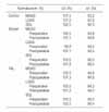
The ankle ROM, knee ROM, maximal muscle length and peak-to-peak excursion during gait improved after surgery. There was a significant difference between the preoperative and postoperative measurements (p<0.05), but no difference between the postoperative and control groups (p>0.05). The muscle length of the soleus was elongated during gait after the Strayer procedure.
Figures and Tables
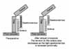
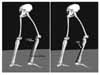 | Fig. 1Muscle modeling using SIMM. Muscle model was made for the lateral head of gastrocnemius (LGAS), medial head of gastrocnemius (MGAS), and soleus (SOL). The changes of muscle lengths during gait were measured with these muscle models.
|
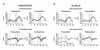
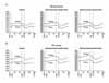 | Fig. 5The changes of muscle length during gait in patient group at preoperative and postoperative gait analysis.
|
References
1. Arnold AS, Delp SL. Computer modeling of gait abnormalities in cerebral palsy: application to treatment planning. Theoretical Issues in Ergonomics Science. 2005. 6:305–312.

2. Chung CY, Ahn CH, Choi IH, Cho TJ, Shin YW, Yoo WJ. Kinematic and kinetic changes of the ankle after the correction of spastic equinus deformity: Z-plastic lengthening versus Strayer method. J Korean Orthop Assoc. 2002. 37:759–765.
3. Delp SL, Loan JP. A graphics-based software system to develop and analyze models of musculoskeletal structures. Comput Biol Med. 1995. 25:21–34.
4. Etnyre B, Chambers CS, Scarborough NH, Cain TE. Preoperative and postoperative assessment of surgical intervention for equinus gait in children with cerebral palsy. J Pediatr Orthop. 1993. 13:24–31.
5. Fish DJ, Nielsen JP. Clinical assessment of human gait. Journal of Prosthetics & Orthotics. 1993. 5:39–48.
6. Gage JR, Fabian D, Hicks R, Tashman S. Pre- and post-operative gait analysis in patients with spastic diplegia: a preliminary report. J Pediatr Orthop. 1984. 4:715–725.
7. Goldstein M, Harper DC. Management of cerebral palsy: equinus gait. Dev Med Child Neurol. 1999. 43:563–569.
8. Hemo Y, Macdessi SJ, Pierce RA, Aiona MD, Sussman MD. Outcome of patients after Achilles tendon lengthening for treatment of idiopathic toe walking. J Pediatr Orthop. 2006. 26:336–340.
9. Lyon R, Liu X, Schwab J, Harris G. Kinematic and kinetic evaluation of the ankle joint before and after tendo achilles lengthening in patients with spastic diplegia. J Pediatr Orthop. 2005. 25:479–483.
10. Perry J, Hoffer MM, Giovan P, Antonelli D, Greenberg R. Gait analysis of the triceps surae in cerebral palsy. A preoperative and postoperative clinical and electromyographic study. J Bone Joint Surg Am. 1974. 56:511–520.
11. Rose SA, DeLuca PA, Davis RB 3rd, Ounpuu S, Gage JR. Kinematic and kinetic evaluation of the ankle after lengthening of the gastrocnemius fascia in children with cerebral palsy. J Pediatr Orthop. 1993. 13:727–732.
12. Joo SY, Park HW, Park KB, Kim HW. Changes in gait pattern after surgeries for equinus gait in cerebral palsy spastic hemiplegia. J Korean Orthop Assoc. 2005. 40:709–716.
13. Winters TF Jr, Gage JR, Hicks R. Gait patterns in spastic hemiplegia in children and young adults. J Bone Joint Surg Am. 1987. 69:437–441.
14. Wren TA, Do KP, Kay RM. Gastrocnemius and soleus lengths in cerebral palsy equinus gait-differences between children with and without static contracture and effects of gastrocnemius recession. J Biomech. 2004. 37:1321–1327.
15. Yngve DA, Chambers C. Vulpius and Z-lengthening. J Pediatr Orthop. 1996. 16:759–764.
 XML Download