

 PDF
PDF ePub
ePub Citation
 Print
Print


Abstract
Osteopetrosis is a very rare hereditary musculoskeletal disorder with an unknown cause. There are few reports on this condition with most focusing on long bone fractures. Most patients with osteopetrosis require non-surgical treatment and surgery is technically difficult. Hallux valgus deformities in patients with osteopetrosis are always severe and there are no reports on its treatment. We treated a hallux valgus deformity using triple osteotomy method and experienced nonunion, subluxation of the first metatarsophalangeal joint and screw breakage. This is the first report of the surgical treatment of such a case in Korea. We report this case with a review of the relevant literature.
Figures and Tables
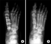
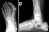 | Fig. 1(A) This preoperative standing anteroposterior radiograph shows highly increased hallux valgus angle, 1st-2nd intermetatarsal angle and distal metatarsal articular angle. (B) In lateral radiograph, bone within a bone in distal tibia and calcaneus is manifested.
|
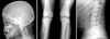 | Fig. 2These radiographs show sclerotic change on skull base, Erlenmyer flask deformity of distal femur and sandwich vertebrae.
|
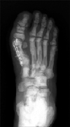
 | Fig. 3This postoperative anteroposterior radiograph shows the correction of hallux valgus angle, intermetatarsal angle and distal metatarsal articular angle.
|
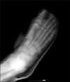
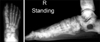
 | Fig. 4At 9 weeks after operation, standing anteroposterior radiograph shows loss of correction and nonunion at all osteotomy sites.
|
 | Fig. 5(A) At postoperative anteroposterior radiograph, distal metatarsal articular angle, hallux valgus angle and intermetatarsal angle were corrected well by using plate and screws. (B) At 30 weeks after reoperation, this anteroposterior radiograph shows complete bony union at osteotomy site, maintenance of corrected angles and breakage of a proximal screw.
|
References
1. Albers-Schonberg H. Roentgenbilder einer seltenen Knochener-Krankung. Munchen Med Wchnschr. 1904. 51:365.
2. Chahabra A, Westlund LA, Kline AJ, McLaughlin R. Management of proximal femoral shaft fractures in osteopetrosis: a case series using internal fixation. Orthopedics. 2005. 28:587–592.

3. Choi WS, Im JI, Kim BH. Open reduction & internal fixation of long bone fracture in osteopetrosis patient. J Korean Fracture Soc. 1995. 8:407–412.
4. Kim ID, Lee SY, Ihin JC, Paik YH. A familial osteopetrosis. J Korean Orthop Assoc. 1976. 11:483–488.
5. Kleinberg S. Osteopetrosis. Am J Surg. 1954. 87:50–62.
6. Milgram JW, Jasty M. Osteopetrosis. A morphological study of twenty-one cases. J Bone Joint Surg Am. 1982. 64:912–929.
7. Mitchell DC. Fractures in brittle bone diseases. Orthop Clin North Am. 1972. 3:787–792.
8. Montgomery RD, Standard KL. Albers-Schonberg's disease. A changing concept. J Bone Joint Surg Br. 1960. 42:303–312.
9. Tachdjian MO. Pediatric Orthopedics. 1990. 2nd ed. Philadelphia: WB Saunders Co;112–115.
10. Yune SH, Rhee KJ, Ahn SR, Kim HY. Osteopetrosis. J Korean Orthop Assoc. 1981. 16:467–470.
 XML Download