

 PDF
PDF ePub
ePub Citation
 Print
Print


Abstract
Purpose
Posterior tibialis tendon dysfunction (PTTD) is one of the most common causes of acquired flatfoot deformity in western countries. But it was known that they were very rare in eastern countries. So we want to report the clinical features and outcomes of 12 patients with PTTD.
Materials and Methods
We evaluated the results of 12 patients using clinical features and results from March 2000 to January 2007 and mean follow up periods is 32 months.
Results
Average age was 45 years, 8 of 12 patients were female, 2 patient with hypertension and 1 with rheumatoid arthritis. 5 patients were overweighted and 5 patients were obese. 4 patients has a history of last trauma. 4 patients experienced progression of flatfoot. On behalf of Johnson and Strom classifications 6 cases were grade I, 4 cases were grade II, grade III, and grade IV was 1 case, relatively. As a treatment we used tenosynovectomy for 3 cases of grade I, additional FDL transfer was done for 2 cases of grade I and 2 cases of grade II. For other 2 patients of grade II flexor digitorum longus transfer and Medial displacement calcaneal osteotomy was done. Pathologic findings of tendon showed degenerative tendinitis. Lastly conservative treatment group was 3 cases of grade I, III, IV each. Average preoperative and postoperative American Orthopedic Foot and Ankle Society's hindfoot/ankle scoreFAS score was 58 and 90. Initial and follow up AOFAS scores of the conservative group was 38 and 57, relatively.
Figures and Tables
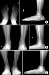
 | Fig. 1Staged plain standing radiogram of PTTD. As the stages rise, increase in talonavicular coverage angle and talocalcaneal angle appear on anteroposterior (AP) radiogram, and talus sagging on lateral radiogram. (A) Johnson and Strom classification grade II: Increased talonavicular coverage angle on anteroposterior radiogram and sagging of talus on lateral radiogram. (B) Johnson and Strom classification grade III: Increased sagging of talus on ankle lateral standing radiogram, and fibulo-calcaneal impingement on both ankle anteroposterior standing radiogram. (C) Johnson and Strom classification grade IV: Valgus tilt of talus, decreased total height of the ankle and fibulocalcaneal impingement of left ankle on both ankle standing anteroposterior radiogram. Note the increased sagging of the talus on standing lateral radiogram.
|
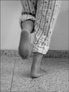
 | Fig. 2Single-limb heel raise test: The patient with posterior tibial tendon dysfunction with flatfoot on the right is being tested for strength of the posterior tibial tendon and muscle with a single heel rise test.
|
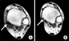 | Fig. 3Magnetic resonance imaging scan shows delayed rupture of posterior tibial tendon. (A) Initial MRI represented synovitis of posterior tibial tendon but, the continuity was intact (arrow). (B) Follow up MRI of 5 months later showed loss of posterior tibial tendon continuity (arrow).
|
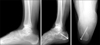 | Fig. 4Preoperative and postoperative ankle lateral radiographs of a Johnson and Strom classification grade II PTTD: Preoperative and postoperative radiographs of the FDL transfer and medial calcaneal displacement osteotomy.
|
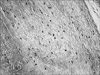 | Fig. 5Histologic appearances of posterior tibialis tendon. Tendon shows fibroblastic proliferation, neovascularization and mucinous degeneration. A few scattered lymphocytes are noted (Hematoxilin and Eosin stain, ×200).
|
Table 1
Summary of the Cases
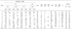
$/¥, Preoperative/Postoperative; *, Talonavicular agle; †, Talo-1stMT angle; ‡, Talocalcaneal angle; ※, Johnson & Strom Classification; ₣, Tenosynovectomy and Debridement; ₤, FDL transfer with MDCO (Medial Displacement Calcaneal Osteotomy); ₱, conservative Tx; €,FDL transfer with Synovectomy and Debridement.
![]()
References
1. Chao W, Wapner KL, Lee TH, Adams J, Hecht PJ. Nonoperative management of posterior tibial tendon dysfunction. Foot Ankle Int. 1996. 17:736–741.

2. Churchill RS, Sferra JJ. Posterior tibial tendon insufficiency. Its diagnosis, management and treatment. Am J Orthop. 1998. 27:339–347.
3. Clain MR, Baxter DE. Simultaneous calcaneocuboid and talonavicular fusion. Long-term follow-up study. J Bone Joint Surg Br. 1994. 76:133–136.
4. Conti S, Michelson J, Jahss M. Clinical significance of magnrtic resonance imaging in preoperative planning for reconstruction of posterior tibial tendon ruptures. Foot Ankle. 1992. 13:208–214.
5. Deland JT, Arnoczky SP, Thompson FM. Adult acquired flatfoot deformity at the talonavicular joint: reconstruction of the spring ligament in an in vitro model. Foot Ankle. 1992. 13:327–332.
6. Duncan JW, Lovell WW. Modified Hoke-Miller flatfoot procedure. Clin Orthop Relat Res. 1983. 181:24–27.
7. Dyal CM, Feder J, Deland JT, Thompson FM. Pes planus in patients with posterior tibial tendon insufficiency: asymptomatic versus symptomatic foot. Foot Ankle Int. 1997. 18:85–88.
8. Evans D. Calcaneo-Valgus deformity. J Bone Joint Surg Br. 1975. 57:270–278.
9. Faciszewski T, Burks RT, Manaster BJ. Subtle injuries of the lisfranc joint. J Bone Joint Surg Am. 1990. 72:1519–1522.
10. Ghormley RK, Spear IM. Anomalies of the posterior tibial tendon a cause of persistent pain about the ankle. AMA Arch Surg. 1953. 66:512–516.
11. Graves SC, Mann RA, Graves KO. Triple arthrodesis in older adults. Results after long-term follow-up. J Bone Joint Surg Am. 1993. 75:355–362.
12. Holmes GB Jr, Mann RA. Possible epidemiological factors associated with rupture of the posterior tibial tendon. Foot Ankle. 1992. 2:70–79.
13. Jahss MH. Disorders of the Foot and Ankle. 1991. 2nd ed. Philadelphia: WB Saunders;1461–1513.
14. Johnson J, Harris G. Pathomechanics of posterior tibial tendon insufficiency. Foot Ankle Clin. 1997. 2:227–239.
15. Johnson KA. Tibialis posterior tendon rupture. Clin Orthop Relat Res. 1983. 177:140–147.
16. Johnson KA, Strom DE. Tibialis posterior tendon dysfunction. Clin Orthop Relat Res. 1989. 239:196–206.
17. Karasick D, Schweitzer ME. Tear of the posterior tibial tendon causing asymmetric flatfoot: radiologic findings. AJR Am J Roentgenol. 1993. 161:1237–1240.
18. Lawrence SJ, Wright RD. Posterior tibial tendon dysfunction: current concepts including operative and nonoperative approaches. Curr Opin Orthop. 2004. 15:62–68.
19. Mann RA. Surgery of the foot and ankle. 1993. 6th ed. St. Lous: C.V.Mosby;757–784.
20. Mann RA, Thompson FM. Rupture of the posterior tibial tendon causing flatfoot. J Bone Joint Surg Am. 1985. 67:556–561.
21. Mays PK, Bishop JE, Laurent GJ. Age-related changes in the proportion of type I and III collagen. Mech Ageing Dev. 1988. 45:203–212.
22. Miller SD, Van Holsbeeck M, Boruta PM, Wu KK, Katcherian DA. Ultrasound in the diagnosis of posterior tibial tendon pathology. Foot Ankle Int. 1996. 17:555–558.
23. Moister SM, Lucas DR, Pomeroy G, Manoli A 2nd. Pathology of the posterior tibial tendon in posterior tibial tendon insufficiency. Foot Ankle Int. 1998. 19:520–524.
24. Myerson MS, Corrigan J, Thompson F, Schon LC. Tendon transfer combined with calcaneal osteotomy for treatment of posterior tibial tendon insufficiency: a radiological investigation. Foot Ankle Int. 1995. 16:712–718.
25. Myerson MS. Adult acquired flatfoot deformity. Treatment of dysfunction of the posterior tibial tendon. J Bone Joint Surg Am. 1997. 78:1434.
26. Myerson M, Solomon G, Shereff M. Posterior tibial tendon dysfunction: its association with seronegative inflammatory disease. Foot Ankle. 1989. 9:219–225.
27. Rose GK. Jahss MH, editor. Pes planus. Disorders of the foot and ankle:medical and surgical management. 1991. 2nd ed. Philadelphia: WB Saunders;892–920. Vol 1.
28. Sangeorzan BJ, Mosca V, Hansen ST Jr. Effect of calcaneal lengthening on relationships among the hindfoot, midfoot, and forefoot. Foot Ankle. 1993. 14:136–141.
29. Schweitzer ME, Caccese R, Karsick D, Wapner KL, Mitchell DG. Posterior tibial tendon tears: utility secondary signs for MR imaging diagnosis. Radiology. 1993. 188:655–659.
30. Sferra JJ, Rosenberg GA. Nonoperative treatment of posterior tibial tendon pathology. Foot Ankle Clin. 1997. 2:261–273.
31. Slovenkai MP. Adult flatfoot: posterior tibial tendon dysfunction, clinical and radiographic evaluation. Foot Ankle Clin. 1997. 2:241–260.
32. Teasdall RD, Johnson KA. Surgical treatment of stage I posterior tibial tendon dysfunction. Foot Ankle Int. 1994. 15:646–648.
33. Wapner KL, Chao W. Nonoperative treatment of posterior tibial tendon dysfunction. Clin Orthop Relat Res. 1999. 365:39–45.
 XML Download