

 PDF
PDF ePub
ePub Citation
 Print
Print


Abstract
Purpose
Phocomelia is an extremely rare congenital anomaly of the upper extremity. There have been no clinical reports about phocomlia in Korea except for five birth reports. We present here the clinical features, classifications and surgical treatments of our phocomelia cases.
Materials and Methods
From January 1993 to August 2007, seven patients were diagnosed as having phocomelia in 9 upper extremities at our clinic. Surgical treatments were performed for five patients on their hand anomalies. We retrospectively reviewed the medical records and radiographs of our cases, and we tried to classify them by the previously suggested systems. We evaluated the functional improvement and measured the VAS scale for parental satisfaction with the operative outcomes.
Results
We could not find any problems during the fetal periods or any hereditary features. The bilaterally-affected patients also had deformities of the lower extremity, while the unilaterally-affected patients did not. We couldn't classify our cases according to the Frantz and O'Rahilly system. We found that the classifications suggested by Tytherleigh-Strong and Hooper (2003) and Goldfarb et al. (2005) could be promising alternatives for classification. One upper extremity was classified as type A, one as type B, and 7 as type C by Tytherleigh-Strong and Hooper's system. Using the Goldfarb's system, two upper extremities were classified as proximal radial longitudinal dysplasia, and seven were classified as proximal ulnar longitudinal dysplasia. Three patients who underwent pollicization showed opposition and tip pinch. Two patients who underwent syndactyly division could do lateral pinch. The VAS scale for parental satisfaction with the functional improvement averaged 8.2 postoperatively.
Figures and Tables
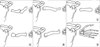
 | Fig. 1The schematic drawings about three types of phocomelia described by Frantz and O'Rahillly. Type I (A), Type II (B) and Type III (C).
|
 | Fig. 2The schematic drawings about three types added by Tytherleigh-Strong and Hooper. Type A (A), Type B (B) and Type C (C).
|
 | Fig. 3The schematic drawings about 3 groups categorized by Goldfarb et al. Proximal radial longitudinal dysplasia (A), Proximal ulnar longitudinal dysplasia (B), Severe combined dysplasia type A (C), Severe combined dysplasia type B (D).
|
 | Fig. 4Unilateral phocomelia (Rt.) with intact shoulder joint (Case 4) X-ray shows relatively intact right shoulder joint. (A) There is only one forearm bone, to which humerus was fused. (B) Hand has only two digits as syndactyly. (C) This case can be classified as type C by Tytherleigh-Strong and Hooper and proximal radial longitudinal dysplasia by Goldfarb et al.
|
 | Fig. 5Unilateral phocomelia (Lt.) with contralateral upper extremity deformity. (Case 2) Lt. upper extremity radiograph shows abnormal glenoid, absence of proximal humerus and synostosis of ulna and radius. (A) Lt. hand had 4 digits with syndactyly of 1st and 2nd finger (B). Rt. upper extremity has radial club hand and hypoplastic thumb. (C) This case can be classified as type B by Tytherleigh-Strong and Hooper and proximal radial longitudinal dysplasia by Goldfarb et al.
|
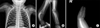
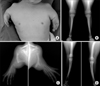 | Fig. 6Bilateral phocomelia. (Case 7) Radiographs shows abnormal shoulder joint, single arm bone with medial exostosis and hand with 5-digits. (C) Bilateral fibular hemimelia was accompanied. (B & D) Note symmetric deformities of the both upper extremity and lower extremity. This case can be classified as type C by Tytherleigh-Strong and Hooper and proximal ulnar longitudinal dysplasia by Goldfarb et al.
|
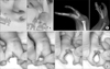 | Fig. 7A case with pollicization and web space deepening. (Case 5) Preoperative photos and x-rays show only 2 digit, which cannot be opposed each other. (A) Pollicization was done with metacarpal shortening and pronation. (B) Intraoperative photos showing possible opposition, palmar view (C) and dorsal view (D).
|
References
1. Baek GH, Chung MS, Park YB, Yoo KH. The relative incidence of congenital anomalies of the hand. J Korean Orthop Assoc. 1997. 32:796–801.

2. Chung MS, Yoon JO, Lee FY. A functional classification of congenital anomalies of the hand. J Korean Orthop Assoc. 1988. 3:109–118.
3. Chung MS, Baek GH, Gong HS. Chung MS, editor. Congenital anomaly. Hand surgery. 2005. 1st ed. Seoul: The Koonja Publishing Inc;1397–1398.
4. Flatt AE. The care of congenital hand anomalies. 1977. St. Louis: The CV Mosby Co;37–51.
5. Fletcher I. Review of the treatment of thalidomide children with limb deficiency in Great Britain. Clin Orthop Relat Res. 1980. 148:18–25.
6. Frantz CH, O'Rahilly R. Congenital skeletal limb deficiencies. J Bone Joint Surg. 1961. 43:1202–1224.
7. Goldfarb CA, Manske PR, Busa R, Mills J, Carter P, Ezaki M. Upper-extremity phocomelia reexamined: a longitudinal dysplasia. J Bone Joint Surg Am. 2005. 87:2639–2648.
8. Hur EJ, Park HM, Kim WK, Bae DW. A case of phocomelia. Korean J Obstet Gynecol. 1987. 30:1427–1433.
9. Johnson HA. Formation of a functional thumb post with sensation in phocomelia. J Bone Joint Surg Am. 1967. 49:327–332.
10. Kim YP, Kim BJ, Chung SR, Lee HJ. A case of phocomelia. Korean J Obstet Gynecol. 1986. 29:282–286.
11. Lamb DW, MacNaughtan AK, Fragiadakis EG. Phocomelia of the upper limb. Hand. 1971. 3:200–203.
12. Lee HJ, Kim DH, Kim HC, IM YG. A case of congenital phocomelia. Korean J Obstet Gynecol. 1973. 16:33–35.
13. Lee SJ, Chai JS, Chang HY, Bae DW. Two cases of phocomelia and chondrodystrophia fetalis. Korean J Obstet Gynecol. 1974. 17:339–342.
14. Shimizu J, Suruga K, Takano S. Therapy of phocomelia and the results of Sulamaa's operation. Seikei Geka. 1967. 18:304–307.
15. Sulamaa M. Upper extremity phocomelia. A contribution to its operative treatment. Clin Pediatr (Phila). 1963. 2:251–257.
16. Sulamaa M, Ryoppy S. Early treatment of congenital bone defects of the extremities: aftermath of thalidomide disaster. Lancet. 1964. 18:130–132.
17. Taussig HB. A study of the German outbreak of phocomelila. The thalidomide syndrome. JAMA. 1962. 180:1106–1114.
18. Taussig HB. Thalidomide and phocomelia. Pediatrics. 1962. 30:654–659.
19. Tytherleigh-Strong G, Hooper G. The classification of phocomelia. J Hand Surg [Br]. 2003. 28:215–217.
20. Whang KH, Kim CH, Lee SJ, Lee SJ. A case of atypical phocomelia. Korean J Obstet Gynecol. 1967. 10:525–528.
21. Yamauchi Y, Nakamura S, Suzuki K, Suruga K. Sulamaa's operation for phocomelia; 7-year follow-up result. Seikei Geka. 1971. 22:907–908.
 XML Download