

 PDF
PDF ePub
ePub Citation
 Print
Print


Abstract
Purpose
It is very difficult to measure the spinal canal dimension in elderly patients because of disc degeneration and facet joint hypertrophy. The purpose of this study is to determine reference values of the spinal canal dimension in a population of normal Korea subjects and to evaluate other measurement methods of the spinal canal dimension that correlate to normal spinal canal dimensions determined using Magnetic Resonance Imaging (MRI).
Materials and Methods
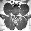
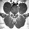
We studied 100 patients who had mild symptoms and had normal MRI findings from 2475 outpatients that had undergone lumbar MRI from November 2002 to May 2004. The dimension of the spinal canal and dural sac was measured at the center of intervertebral discs L3/4, L4/5 and L5/S1. The dimension of the spinal canal and vertebral body was measured and was compared at the transverse plane perpendicular to the spinal canal that transected L4, L5 and the S1 pedicle.
Results
For the sequence of L3/4, L4/5 and L5/S1, the mean spinal canal dimensions were 249.38±38.30 mm2, 253.04±48.62 mm2 and 288.46±57.62 mm2, respectively. For the sequence of L4, L5 and S1, the mean spinal canal dimensions were 279.78±42.36 mm2, 301.50±54.26 mm2 and 355.10±60.65 mm2, respectively. The correlation coefficient was high at 0.913 for the L3/4 and L4 interpedicular transverse plane. The correlation coefficient for L4/5 and L5 was 0.905, and the correlation coefficient for L5/S1 and S1 was 0.845.
Conclusion
The lumbar spinal canal dimension measured at the intervertebral disc level in a population of normal Korean subjects is expected to be useful as reference data. The transpedicular plane perpendicular to the spinal canal can give information for estimating the spinal canal dimension at the disc level.
Figures and Tables


References
1. Arnoldi CC, Brodsky AE, Cauchoix J, et al. Lumbar spinal stenosis and nerve root entrapment syndromes. Definition and classification. Clin Orthop Relat Ress. 1976. 115:4–5.
2. Becker SH, Arenson RL. Costs benefits of picture archiving and communication systems. J Am Med Inform Assoc. 1994. 1:361–371.
3. Clark K. Significance of the small lumbar spinal canal: cauda equina compression syndromes due to spondylosis. 2. Clinical and surgical significance. J Neurosurg. 1969. 31:495–498.
4. Dora C, Wälchli B, Elfering A, Gal I, Weishaupt D, Boos N. The significance of spinal canal dimensions in discriminating symptomatic from asymptomatic disc herniations. Eur Spine J. 2002. 11:575–581.

5. Elsberg CA, Dyke CG. Diagnosis and localization of tumors of spinal cord by means of measurements made on X-ray films of vertebrae and the correlation of clinical and X-ray findings. Bull Neurol Inst NY. 1934. 3:359–375.
6. Epstein JA, Epstein BS, Lavin L. Nerve root compression associated with narrowing of the lumbar spinal canal. J Neurol Neurosurg Psychiatry. 1962. 25:165–176.
7. Frymoyer JW. Back pain and sciatica. N Engl J Med. 1988. 318:291–300.
8. Gill GG, White H. Mechanisms of nerve-root compression and irritation in backache; surgical decompression in intervertebral disk conditions; spondylolisthesis, spina bifida occulta and transitional fifth lumbar vertebra. Clin Orthop Relat Res. 1955. 5:66–81.
9. Haughton VM. MR imaging of the spine. Radiology. 1988. 166:297–301.
10. Masaryk TJ, Ross JS, Modie MT, Boumphrey F, Bohlman H, Wilber G. High-Resolution MR imaging of sequestered lumbar intervertebral disks. AJR Am J Roentgenol. 1988. 150:1155–1162.
11. Modic MT, Pavlicek W, Weinstein MA, et al. Magnetic resonance imaging of intervertebral disk disease. Clinical and pulse sequence considerations. Radiology. 1984. 152:103–111.
12. Porter RW, Ottowell D, Wicks M. Use of diagnostic ultrasound for spinal canal measurements. J Bone Joint Surg Br. 1977. 59:249–250.
13. Verbiest H. A radicular syndrome from developmental narrowing of the lumbar vertebral canal. J Bone Joint Surg Br. 1954. 36:230–237.
14. Verbiest H. Further experiences on the pathological influence of a developmental narrowness of bony lumbar vertebral canal. J Bone Joint Surg Br. 1955. 37:576–583.
15. Verbiest H. Results of surgical treatment of idiopathic developmental stenosis of the lumbar vertebral canal. A review of twenty-seven years' experience. J Bone Joint Surg Br. 1977. 59:181–188.
16. Winston K, Rumbaugh C, Colucci V. The vertebral canals in lumbar disc disease. Spine. 1984. 9:414–417.
 XML Download