

 PDF
PDF ePub
ePub Citation
 Print
Print


Abstract
Purpose
In this study, we investigated whether thoracoscopic anterior correction can effectively derotates the scoliotic spine and we also evaluated the patterns of derotational changes inside the instrumented area and at the junction between the instrumented and uninstrumented area.
Materials and Methods
Preoperative and postoperative MR images with single axial cuts through each vertebral level were obtained in 20 patients who underwent thoracoscopic anterior instrumentation. Each vertebral rotation was measured by the use of Aaro's method. Vertebral axial derotation of the apical zone (AZ), upper instrumented zone (UIZ), lower instrumented zone (LIZ) and junctional segment were compared. The amount of segmental rotation and segmental derotation in each zone was calculated. Statistical analysis was performed by the use of by 2-way ANOVA and the Wilcoxon signed ranks test.
Results
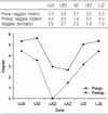
The average axial rotations at AZ were 10.1° preoperatively and 5.3° postoperatively with an average correction rate of 48%. Derotation of the AZ was greater than the UIZ and LIZ. For the LIZ derotation was not significant (p=0.023) while for the UIZ there was a significant derotation (p<0.001). Postoperatively, the first uninstrumented vertebra rotated significantly in the direction of rotation of the instrumented end vertebra. Preoperative and postoperative segmental rotation was higher in the UIZ and LIZ than in the AZ. However, segmental derotation occurred uniformly in the instrumented area. At the junctional segment, preoperative segmental rotation was same as that of the UIZ and LIZ and segmental derotation was not significant.
Figures and Tables
Fig. 1
MRI scans of axial slices through each vertebral level (A). Vertebral rotation (RA sag) measures the angle between the long axis of the vertebra subtended at the posterior central aspect of the spinal canal, and the sagittal plane (B).
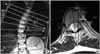
Fig. 2
Definitions of zone and segment. The apical zone was defined as the apical vertebra and the vertebra above and below it. The upper instrumented zone consisted of the two most cranial vertebra within the instrumentation and the lower instrumented zone consisted of the two most caudal vertebrae within the instrumentation. The segment just outside of instrumentation was defined as the upper and lower junctionnal segment. UEV, upper end vertebra; AV, apical vertebra; LEV, lower end vertebra.
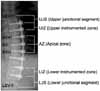
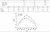
Table 2
Changes of Segmental Rotation (Degree)
UJZ, upper junctional zone; UEZ, upper end vertebral zone; AZ, apical zone; LEZ, lower end vertebral zone; LJZ, lower junctional zone; UJS, upper junctional segment; UIZ, upper-instrumented zone; UAZ, upper apical zone, LAZ, lower apical zone; LIZ, lower instrumented zone; LJS, lower junctional segment.
References
1. Aaro S, Dahlborn M, Svensson L. Estimation of vertebral rotation in structural scoliosis by computer tomography. Acta Radiol Diagn (Stockh). 1978. 19:990–992.

2. Betz RR, Harms J, Clements DH 3rd, et al. Comparison of anterior and posterior instrumentation for correction of adolescent thoracic idiopathic scoliosis. Spine. 1999. 24:225–239.
3. Kaneda K, Shono Y, Satoh S, Abumi K. Anterior correction of thoracic scoliosis with Kaneda anterior spinal system. A preliminary report. Spine. 1997. 22:1358–1368.
4. Lee SM, Suk SI, Chung ER. Direct vertebral rotation: a new technique of three-dimensional deformity correction with segmental pedicle screw fixation in adolescent idiopathic scoliosis. Spine. 2004. 29:343–349.
5. Picetti GD 3rd, Ertl JP, Bueff HU. Endoscopic instrumentation, correction, and fusion of idiopathic scoliosis. Spine J. 2001. 1:190–197.
6. Wood KB, Olsewski JM, Schendel MJ, Boachie-Adjei O, Gupta M. Rotational changes of the vertebral pelvic axis after sublaminar instrumentation in adolescent idiopathic scoliosis. Spine. 1997. 22:51–57.
7. Wood KB, Transfeldt EE, Ogilvie JW, Schendel MJ, Bradford DS. Rotational changes of the vertebral-pelvic axis following Cotrel-Dubousset instrumentation. Spine. 1991. 16:Suppl 8. S404–S408.
8. Zielke K. Ventral derotation spondylodesis. Results of treatment of cases of idiopathic lumbar scoliosis (author's (author's transl)). Z Orthop Ihre Grenzgeb. 1982. 120:320–329.
 XML Download