

 PDF
PDF ePub
ePub Citation
 Print
Print


Abstract
Although a closing wedge high tibial osteotomy can decrease the posterior tibial slope, there are no reports of severe genu recurvatum after a closing wedge osteotomy that required corrective surgery. A 54-year-old male with medial compartment osteoarthritis and genu varum was treated with a closing wedge high tibial osteotomy in another hospital, which led to severe recurvatum because of surgical failure. He complained of knee pain and a severe deformity, but of which were corrected by an open wedge corrective osteotomy, a two-wedge bicortical autograft reconstruction, and double plate fixation. Surgeons should pay close attention to both varus deformity correction and changes in the posterior tibial slope during a closing wedge high tibial osteotomy.
Figures and Tables
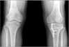
Fig. 1
Preoperative radiographs of a 54-year-old male showing genu varum with medial compartment osteoarthritis. The femorotibial angle was varus 5 degree and the tibial posterior slope was 7 degrees.
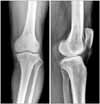
Fig. 2
Three months after the closing wedge osteotomy in another hospital, postoperative radiographs show the correction of genu varum but severe genu recurvatum. The femorotibial angle was valgus 2 degrees and the tibial posterior slope was -15 degrees.
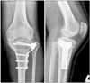
Fig. 3
Ten months after the closing wedge osteotomy, radiographs show persistent genu recurvatum and loss of valgus correction. The femorotibial angle was varus 2 degrees and the tibial posterior slope was -14 degrees.
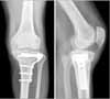
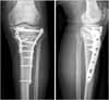
Fig. 4
Four months after the corrective osteotomy, radiographs show the correction of genu recurvatum and genu varum. The femorotibial angle was valgus 5 degrees and the tibial posterior slope was 4 degrees.
References
1. Chen LC, Chan YS, Wang CJ. Opening-wedge osteotomy, allografting with dual buttress plate fixation for severe genu recurvatum caused by partial growth arrest of the proximal tibial physis: a case report. J Orthop Trauma. 2004. 18:384–387.
2. Coventry MB. Osteotomy about the knee for degenerative and rheumatoid arthiritis. J Bone Joint Surg Am. 1973. 55:23–48.
3. Jackson JP. Osteotomy for osteoarthritis of the knee, in proceedings of the sheffield regional orthopedic club. J Bone Joint Surg. 1958. 40:826.
4. Lee JY, Seon JK, Song EK, Yoon TR, Cheon SY, Lim KY. Comparison of high tibial osteotomy - opening versus closing wedge osteotomy -. J Korean Orthop Assoc. 2004. 39:790–796.
5. Marti CB, Gautier E, Wachtl SW, Jakob RP. Accuracy of frontal and sagittal plane correction in open-wedge high tibial osteotomy. Arthroscopy. 2004. 20:366–372.

6. Moroni A, Pezzuto V, Pompili M, Zinghi G. Proximal osteotomy of the tibia for the treatment of genu recurvatum in adults. J Bone Joint Surg Am. 1992. 74:577–586.
7. Poilvache P. Scott WN, editor. Osteotomy for the arthritic knee: a European perspective. Surgery of the knee. 2006. 4th ed. Philadelphia: Elsevier Inc;1321–1366.
8. Sundaram NA, Hallett JP, Sullivan MF. Dome osteotomy of the tibia for osteoarthritis of the knee. J Bone Joint Surg Br. 1986. 68:782–786.
9. Vail TP, Lang JE. Scott WN, editor. Surgical techniques and instrumentation in total knee arthroplasty. Surgery of the knee. 2006. 4th ed. Philadelphia: Elsevier Inc;1507–1508.
10. Winsor RE, Insall JN, Vince KG. Technical considerations of the total knee arthroplasty after proximal tibial osteotomy. J Bone Joint Surg Am. 1988. 70:547–555.
 XML Download