

 PDF
PDF ePub
ePub Citation
 Print
Print


Primary solitary amyloidosis, or amyloidoma, is a rare subset of amyloidosis in which the amyloid deposition is focal and not secondary to a systemic process or plasma cell dyscrasia9). Amyloidoma in the soft tissue is exceedingly rare and occurs most often in the mediastinum or abdomen6). We report a case of amyloidoma presenting in the tibialis anterior muscle with the radiological features suggesting chronic osteomyelitis.
CASE REPORT
A 44-year-old man presented with pus discharge in the middle of his right leg with three months duration. He had a history of fractures in the right proximal tibia and fibula from a traffic accident 27 years ago and peroneal nerve palsy since then. Eight years ago, he experienced painful swelling in the right leg, which improved after taking some medication at a local clinic without a precise diagnosis.
In the middle of the right leg, there were two ulcerative skin lesions 8 and 30mm in diameter, respectively, with some pus discharge. There was a hard mass without tenderness and a clear margin under the skin ulcer with decreased sensory perception in peroneal nerve innervation. The grade of the extensor hallusis and anterior tibial muscles was 0 and 1-2, respectively. There was no motion limitation in the knee joint.
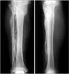
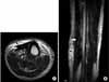
The plain radiograph showed a large amount of high density material in front of the tibia and fibula (Fig. 1). Magnetic resonance imaging of the right leg revealed a densely calcified portion of the mass demonstrating low signal intensity on T2-weighted views, with high signal intensity suggesting fluid (Fig. 2). In addition, there was a moderate amount of enhancement of the noncalcified portions of the mass on the gadopentetate dimeglumine-enhanced T1-weighted views.
Surgical exploration was performed under the impression of chronic osteomyelitis. There was a large amount of sawdust like ossified material scattered along the anterior tibialis muscle (Fig. 3). The mass was evacuated completely. Multiple specimens of the lesion involving the ossified material and soft tissue surrounding it were taken and a pathologic examination and germiculture was undertaken.
Microscopically, the tissue appeared as an amorphous, eosinophilic, hyaline, extracellular substance with extensive calcification and degeneration (Fig. 4A). Congo red staining revealed pink-red deposits of amyloid in most of the tissue obtained. Under polarized light, the Congo red-stained amyloid showed a green birefringence (Fig. 4B). The patient recovered quickly after surgery, and a satisfactory clinical result was achieved at the final follow-up.
DISCUSSION
Amyloidosis is an uncommon disease, in which various organs are infiltrated by amorphous extracellular eosinophilic material composed of insoluble proteins with a high degree of beta-pleated sheet protein structure7). It is a heterogeneous disorder with various manifestations; the most common being systemic1). Amyloid deposits can be found throughout the body in single or multiple organs. A single soft-amyloidoma, as a local amyloidosis, has been reported in widely various anatomic sites, including the respiratory tract, mediastinum, gastrointestinal tract, mesentery, retroperitoneum, genitourinary tract, breast, central nervous system, bone, lymph node, spleen, parotid gland, cornea, conjunctiva, and orbit5,8).
The cause of amyloidosis is unknown. Some investigators believe that amyloid deposition stems from a derangement in immunoregulation after a protracted antigenic challenge that causes an immunoglobulin precursor protein to polymerize in the extracellular space, producing amyloid3). Others believe that amyloid tumors arise from isolated clones of plasma cells, which are producers of immunoglobulin light chains that form amyloid fibrils in the connective tissues through enzymatic degradation7). Others have suggested that localized amyloidosis can arise from burnt-out extramedullary plasmacytomas4). Our patient had a history of a proximal tibia fracture, and the cause of amyloidoma might be associated with the bone and soft tissue injury.
Because soft tissue amyloidoma of the extremity is quite rare, the clinical suspicion of amyloidosis as the cause of an extremity mass is likely to be very low, particularly in the absence of immunocyte dyscrasia.
Although amyloidoma usually presents as lytic lesions, ossification and/or calcification is observed in 29% of the nodules with amyloidosis10). Therefore, the densely calcified mass observed in this case is not an unusual appearance. The mass showed radiographically detectable calcification in the plain radiographic, one with scattered high signals. Coby MJ et al.2) described the MR signal characteristics of amyloid deposition as being intermediate between those of the fibrocartilage and muscle on all sequences. In this study, the mass of amyloidoma showed low (lower than muscle) signal in the T2-weighted images, as described previously, and exhibited moderate enhancement of the signal in the enhanced T1-weighted views. However, because the amyloidoma was accompanied by an infection, there was high signal intensity in the T2-weighted images suggesting the disease to be chronic osteomyelitis.
The imaging findings in amyloidoma are not specific. Therefore, a tissue biopsy is essential for a diagnosis. Amyloid typically stains with Congo red and produces a characteristic green birefringence and dichroism under polarized light. In our case, after the Congo red-stain, a green birefringence was observed under polarized light that demonstrated the lesion to be an amyloid deposit.
Localized amyoidosis without evidence of any systemic disease is a benign lesion that may be treated surgically with a low recurrence rate. Complete resection of the lesion was carried out for our patient and no sign of recurrence was found in the latest follow-up 5 years after surgery.
In conclusion, soft tissue amyloidoma is a rare subtype of amyloidosis that requires a tissue biopsy for a diagnosis. However, it has an excellent prognosis with a local resection.


 XML Download