

 PDF
PDF ePub
ePub Citation
 Print
Print


Abstract
Purpose
This article aims at evaluating the results of treatment which excise the intradural extramedullary tumor using surgical microscope, mainly concerned by the field of orthopaedic surgery.
Materials and Methods
A retrospective review was carried out on 11 cases who were operated on for the excision of intradural extramedulary tumor in two hospitals from June 2001 to May 2007. Of the 11 cases, there were 3 males, and 8 females with an average age of 62.4 (33-78) years. Average follow-up period is 18.8 (1-78) months. Patients were diagnosed by MRI and pathological diagnosis was analyzed. The clinical evaluation was made by the index of VAS (visual analogue scale) and Cooper-Epstein grade.
Figures and Tables
Fig. 1
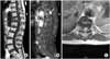
(A) T1-weighted sagittal MR image shows intermediate signal intensity. The tumor is located at the T10-11 level with extension of the tumor forming a dumbbell shaped mass. (B) Gadolinium enhanced T1 weighted sagittal MR image shows well contrast enhancing mass. (C) Gadolinium enhanced T1 weighted axial MR image shows a dumbbell shape intradural extramedullary mass compressing spinal cord to the left.
References
1. Conti P, Pansini G, Mouchaty H, Capuano C, Conti R. Spinal neurinomas: retrospective analysis and long-term outcome of 179 consecutively operated cases and review of the literature. Surg Neurol. 2004. 61:34–43.

2. Cushing H, Eisenhardt L. Meningiomas: their classification, regional behavior, life history, and surgical end results. 1938. 1st ed. Ilinois, Spingfield: Charles C Thomas;735.
3. el-Mahdy W, Kane PJ, Powell MP, Crockard HA. Spinal intradural tumours: part I--extramedullary. Br J Neurosurg. 1999. 13:550–557.
4. Harry N. The spine. 1998. 4th ed. Philadelphia: WB Saunders Co;1366.
5. Jallo GI. CUSA excel ultrasonic aspiration system. Neurosurgery. 2001. 48:695–697.
6. Jinnai T, Koyama T. Clinical characteristics of spinal nerve sheath tumors: analysis of 149 cases. Neurosurgery. 2005. 56:510–515.
7. McCormick PC. Anatomic principles of intradural spinal surgery. Clin Neurosurg. 1994. 41:204–223.
8. Nittner K. Spinal meningiomas, neurinomas, and neurofibroma-haourglass tumors. Handbook of clinical neurology. 1976. 20. Amsterdam: North-Holland Publishing Co;177–322.
9. Roux FX, Nataf F, Pinaudeau M, Borne G, Devaux B, Meder JF. Intraspinal menigiomas: review of 54 cases with discussion fo poor prognosis factors and modern therapeutic management. Surg Neurol. 1996. 46:458–463.
10. Shim DM, Kim SS, Lee BC, Choi ES. Clinical analysis and comparative study of standard and microsurgical lumbar discectomy. J Kor Soc Spine Surg. 1994. 1:87–92.
11. Shin BJ, Lee JC, Yoon TK, et al. Surgical treatments of intradural extramedullary tumor. J Kor Soc Spine Surg. 2002. 9:230–237.
12. Souweidane MM, Benjamin V. Spinal cord meningiomas. Neurosurg Clin N Am. 1994. 5:283–291.
13. Traul DE, Shaffrey ME, Schiff D. Part I: spinal-cord neoplasms-intradural neoplasms. Lancet Oncol. 2007. 8:35–45.
14. Van Goethem JW, van den Hauwe L, Ozsarlak O, De Schepper AM, Parizel PM. Spinal tumors. Eur J Radiol. 2004. 50:159–176.
 XML Download