

 PDF
PDF ePub
ePub Citation
 Print
Print


Abstract
Fibrous dysplasia is a developmental anomaly of bone formation, which is characterized by a replacement of the normal bone and marrow by fibrous tissue as well as small, woven spicules of bone. These lesions may exist in a monostotic or polyostotic form and are generally found in the 4th decade of age or younger with a slightly higher incidence in girls than boys. We report a case of monostotic fibrous dysplasia that was associated with a pathologic fracture and a kyphotic deformity in the cervical spine.
Figures and Tables
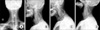
Fig. 1
The plain radiograph shows a ground glass pattern lesion of the body of the 4th and 5th cervical vertebra. (A) Preoperative C-spine AP view. (B) Preoperative C-spine Lateral view. (C) Preoperative C-spine Rt. oblique view. (D) Preoperative C-spine Lt. oblique view.
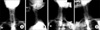
Fig. 2
The computed tomogram shows the loss of the anterior portion of the 4th and 5th cervical vertebra as well as local kyphosis of the cervical spine. (A) Preoperative CT sagittal view. (B) Preoperative C4 axial view. (C) Preoperative C5 axial view.
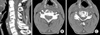
Fig. 3
The preoperative magnetic resonance images shows an expansile and low signal density lesion of the vertebral body, pedicle, lamina and spinal canal involvement of 4th and 5th cervical vertebrae. (A) High signal intensity on the T2-weighted sagittal view. (B) High signal intensity on the T2-weighted C4 axial view. (C) High signal intensity on the T2-weighted C5 axial view.
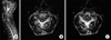
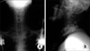
Fig. 4
The histology section shows the vertebrae to be largely replaced by proliferating fibrous connective tissue with focally presenting woven bone trabeculae (chinese letter appearance). (A) H and E stain, ×40. (B) H and E stain, ×100. (C) H and E stain, ×200.

References
1. Dahlin DC. Malignant bone tumors: improvement in prognosis. Mayo Clin Proc. 1988. 63:414–415.

2. Danilu A, Witwicki T. Case of fibrous dysplasia of the spine. Chir Narzadow Ruchu Orthop Pol. 1979. 44:165–167.
3. Harris WH, Dudley HR Jr, Barry RJ. The natural history of fibrous dysplasia. An orthopaedic, pathological and roentgonographic. J Bone Joint Surg Am. 1962. 44:207–233.
4. Kim BJ, KIM YU, Yoo MJ. Monostotic fibrous dysplasia of the cervical spine. J Korean Orthop Assoc. 1988. 23:1221–1226.
5. Lichtenstein L, Jaffe HL. Fibrous dysplasia of bone. A condition affecting one, several or many bones, the graver cases of which may present abnormal pigmentation of skin, premature sexual development, hyperthyroidism or still other extraskeletal abnormalities. Arch Pathol. 1942. 33:77–816.
6. Resnik CS, Lininger JR. Monostotic fibrous dysplasia of the cervical spine: case report. Radiology. 1984. 151:49–50.
7. Rosenblum B, Overby C, Levine M, Hanfler M, Sprecher S. Monostotic fibrous dysplasia of the thoracic spine. Spine. 1987. 12:939–942.
8. Schlumberger HG. Fibrous dysplasia of the single bones (monostotic fibrous dysplasia). Milit Surg. 1946. 99:504–527.
9. Stewart MJ, Gilmer WS, Edmonson AS. Fibrous dysplasia of bone. J Bone Joint Surg Br. 1962. 44:302–318.
10. Weil A. Pubertas praecox und Knochen-bruchigkeit. Klin Wchnschrft. 1932. 2:2114–2115.
 XML Download