

 PDF
PDF ePub
ePub Citation
 Print
Print


Abstract
Purpose
To access the efficacy of a modified design of a reverse superficial sural artery flap (RSSAF) to decrease the level of venous congestion and flap necrosis.
Materials and Methods
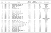
Twenty four cases of RSSAF were performed. The mean age of the patients was 47 years and the mean follow up period was 21 months. The average size of the flap was 7.4×5.2 cm. There were 19 cases of the modified flap design and 5 cases of non-modified design. The venous congestion and complications were analyzed according to the modification of the flap design.
Results
The flap survived in 23 cases. Primary closure of the donor site was performed in 11 cases whose flap width was <5 cm, and 12 cases required a skin graft. Venous congestion was observed in 5 cases including 4 cases, who were not treated with the modified design and 1 case with arterosclerosis obliterans preoperatively. Partial flap necrosis occurred in 2 flaps that did not have the modified design.
Figures and Tables
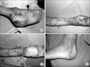 | Fig. 1(A) A 31-year-old man suffered a crushing injury on the heel and foot in a motor vehicle accident. (B) After debridement, there was a 10×6 cm defect, which resulted in exposure of the calcaneal bone on the heel. (C) The defect was covered with a reverse sural artery flap with tear drop skin design on the pedicle. The donor-site wound was managed successfully using a split-thickness skin graft. (D) The functional and cosmetic result was good.
|
 | Fig. 2(A) A 52-year-old man was diagnosed with a malignant melanoma on the heel. After a wide excision, there was a 9×9cm defect. (B) The defect was covered with a reverse sural artery flap with a tear drop skin design on the pedicle. The donor-site defect was covered with a split-thickness skin graft. (C) The final result at 2 years was excellent.
|
References
1. Hasegawa M, Torii S, Katoh H, Esaki S. The distally based superficial sural artery flap. Plast Reconstr Surg. 1994. 93:1012–1020.

2. Hidalgo DA, Shaw WW. Reconstruction of foot injuries. Clin Plast Surg. 1986. 13:663–680.
3. Jeung SF, Wei FC. Distally based sural island flap for foot and ankle reconstruction. Plast Reconstr Surg. 1997. 99:744–750.
4. Mark FP, Peter JC, Paul AW, Salvatore L. Reverse sural artery flap: Caveats for success. Ann Plast Surg. 2002. 48:496–504.
5. Masquelet AC, Romana MC, Wolf G. Skin island flaps supplied by the vascular axis of the sensitive superficial nerves: anatomic study and clinical experiences in the leg. Plast Reconstr Surg. 1992. 89:1115–1121.
6. Oberlin C, Azoulay B, Bhatia A. The posterolateral malleolar flap of the ankle: a distally based neurocutaneous flap--report on 14 cases. Plast Reconstr Surg. 1995. 96:400–407.
7. Ogun TC, Arazi M, Kutlu A. An easy and versatile method of coverage for distal tibial soft tissue defects. J Trauma. 2001. 50:53–59.
8. Rooks MD. Coverage problems of the foot and ankle. Orthop Clin North Am. 1989. 20:723–736.
9. Serkan Y, Mithat A, Tayfun A. Soft-tissue reconstruction of the foot with distally based neurocutaneous flaps in diabetic patients. Ann Plast Surg. 2002. 48:258–264.
10. Tu YK, Steve WN, Yeh WL, Wang KC. Reconstgruction of ankle and heel defects by a modified wide-base reverse sural flap. J Trauma. 1999. 47:82–88.
11. Yilmaz M, Karatas O, Barutcu A. The distally based superficial sural artery island flap: clinical experiences and modifications. Plast Reconstr Surg. 1998. 102:2358–2367.
 XML Download