

 PDF
PDF ePub
ePub Citation
 Print
Print


Abstract
Purpose
The shape of the acromion was analyzed radiologically to assess the effect on rotator cuff tears.
Materials and Methods
The rotator cuff tear group consisted of 91 patients with a mean age 55.6 years (range, 27-85 years) and the normal shoulder articular disease (control) group consisted of 100 subjects with a mean age of 42.9 years (range, 18-72 years). The lateral extension of the acromion and the lateral acromial angle were measured on an oblique coronal MRI, and the anterior covering of the acromionon was measured on a supraspinatus outlet view.
Results
The lateral extension of the acrominon of the rotator cuff tear group of patients and the control group of subjects was 0.70±0.07 and 0.64±0.10, respectively. The lateral acromial angle was 72.6±6.5° and 76.4±6.5°, respectively, and the anterior covering of the acromion was measured to be 0.55±0.13 and 0.51±0.14, respectively. In a comparison with the control group of subjects, the lateral extension of the acrominon of the rotator cuff tear group of patients was larger and the lateral acromial angle was smaller; these findings were statistically significant (p<0.001). In addition, the correlation coefficients of the lateral extension of the acromion to the lateral acromial angle in the rotator cuff tear group of patients and in the control group of subjects was -0.44 and -0.46, respectively; a statistically significant correlation was seeen (p<0.001).
Figures and Tables
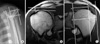 | Fig. 1(A) Measurement of the anterior covering of the acromion from the supraspinatus outlet view. A reference line was drawn parallel to the axis of the humeral diaphysis from the anterior margin of the acromion. The anterior covering of the acromion (A/B) was defined by the ratio of the distance between the posterior margin of the humeral head to this reference line (A) over the distance between the anterior margin to the posterior margin of the humeral head (B). (B) The acromiohumeral distance on the oblique coronal MR image was measured as the shortest distance, in millimeters, between the undersurface of the acromion and the superior edge of the humeral head. (C) The lateral extension of the acromion (A/B) on the oblique coronal MR image was calculated by dividing the distance from the superior and inferior osseous margins of the glenoid cavity to the lateral border of the acromion (A) by the distance from the superior and inferior osseous margins of the glenoid cavity to the most lateral part of the proximal part of the humerus (B).
|
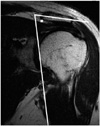 | Fig. 2The lateral acromion angle was measured as the angle between the line through the midsubstance of the acromion. The bony outline of the glenoid cavity on the oblique coronal MR image was measured immediately posterior to the acromioclavicular joint.
|
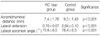
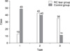 | Fig. 3Radiological comparison between the rotator cuff group of patients and the control group of subjects.
|
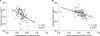 | Fig. 4Correlation between the lateral extension of the acromion and the lateral acromion angle. (A) The rotator cuff tear group of patients; (B) the control group of subjects. *Pearson correlation coefficient, †p-value.
|

References
1. Banas MP, Miller RJ, Totterman S. Relationship between the lateral acromion angle and rotator cuff disease. J Shoulder Elbow Surg. 1995. 4:454–461.

2. Bigliani LU, Morrison DS, April EW. The morphology of the acromion and its relationship to rotator cuff tears. Ortho Trans. 1986. 10:216.
3. Getz JD, Recht MP, Piraino DW, et al. Acromial morphology: relation to sex, age, symmetry, and subacromial enthesophytes. Radiology. 1996. 199:737–742.
4. Loehr J, Uhthoff H. The pathogenesis of degenerative rotator cuff tears. Ortho Trans. 1987. 11:237.
5. MacGillivray JD, Fealy S, Potter HG, O'Brien SJ. Multiplanar analysis of acromion morphology. Am J Sports Med. 1998. 26:836–840.
6. Neer CS 2nd. Anterior acromioplasty for the chronic impingement syndrome in the shoulder. A preliminary report. J Bone Joint Surg Am. 1972. 54:41–50.
7. Neer CS 2nd, Poppen NK. Supraspinatus outlet. Paper presented at ASES 3rd open meeting. 1987. San Francisco.
8. Nyffeler RW, Werner CM, Sukthankar A, Schmid MR, Gerber C. Association of a large lateral extension of the acromion with rotator cuff tears. J Bone Joint Surg Am. 2006. 88:800–805.
9. Ono K, Yamamuro T, Rockwood CA. Use of a thirty-degree caudal tilt radiograph in the shoulder impingement syndrome. J shoulder Elbow Surg. 1992. 1:246–252.
10. Tetreault P, Krueger A, Zurakowski D, Gerber C. Glenoid version and rotator cuff tears. J Orthop Res. 2004. 22:202–207.
11. Toivonen DA, Tuite MJ, Orwin JF. Acromial structure and tears of the rotator cuff. J Shoulder Elbow Surg. 1995. 4:376–383.
12. Tuite MJ, Toivonen DA, Orwin JF, Wright DH. Acromial angle on radiographs of the shoulder: Correlation with the impingement syndrome and rotator cuff tears. AJR Am J Roentgenol. 1995. 165:609–613.
13. Wang JC, Hatch JD, Shapiro MS. Comparison of MRI and radiographs in the evaluation of acromial morphology. Orthopedics. 2000. 23:1269–1271.
14. Zuckerman JD, Kummer FJ, Cuomo F, Rosenblum S, Katz N. The influence of the cor- acoacromial arch anatomy on rotator cuff tears. J Shoulder Elbow Surg. 1992. 1:4–14.
 XML Download