

 PDF
PDF ePub
ePub Citation
 Print
Print


INTRODUCTION
The subacromial bursa is located between the under surface of the coracoacomial arch (CA arch) and the rotator cuff. The bursa and rotator cuff tendon must pass repeatedly underneath the CA arch during shoulder motion. The bursa is believed to have a lubricating effect by reducing the level of friction between the rotator cuff and the CA arch22). However, repeated compression of the rotator cuff tendon under the CA arch can damage the tendon and induce inflammation of the subacromial bursa19). Inflammation in the subacromial bursa can lead to shoulder pain by stimulating the nerve fibers or increasing the levels of neuromediators such as substance P22). Furthermore, in the presence of rotator cuff tears, there may be further inflammation in the subacromial bursa in response to the degenerated tendon tissue23). Despite these proposed mechanisms leading to inflammation of the subacromial bursa, there is no agreement regarding the role of inflammation in subacromial bursitis and rotator cuff disease.
Several studies have implicated the subacromial bursa as a major pathological factor in rotator cuff disease. Soifer et al.27) reported that there was a significantly richer supply of free nerve fibers in the subacromial bursa than in the other tissues; and the nociceptive information relayed by these fibers might be responsible for the pain associated with impingement syndrome. Gotoh et al.8) demonstrated a correlation between an increase in the level of substance P in the subacromial bursa and the pain caused by rotator cuff disease. In previous histology studies, proliferating changes and fibrosis in the synovial tissue of the subacromial bursa were observed but with minimal inflammatory cell infiltration24,25,32). Despite the lack of inflammatory cells, it is believed that inflammatory cytokines are expressed, which stimulate the peripheral nociception without cell infiltration, leading to shoulder pain in rotator cuff disease.
There have been a few reports on the inflammatory cytokines in the subacromial bursa tissue. In rotator cuff disease, the inflammatory response is mediated by complex reactions between cytokines including interleukins (IL-1, IL-6 and IL-8), tumor necrosis factor-α (TNF-α), transforming growth factor (TGF), basic fibroblast growth factor (bFGF) and vascular endothelial growth factor (VEGF)9,23,28,32). However, these cytokines are involved in the inflammatory reaction of the subacromial bursa. To date, there has been no screening study to determine the presence of inflammatory cytokines in subacromial bursitis. Until recently, functional genetic studies have had limited scope and are only able to elucidate the role of one or a few genes at one time. The cDNA Array technique allows the expression of many genes to be monitored simultaneously. The power of this technique lies in its potential for comparative expression studies in diseased versus normal samples15).
The aim of this study was to obtain an overview of the gene expression pattern of inflammatory cytokines/receptors of the subacromial bursa tissue derived from patients with rotator cuff disease compared with normal controls.
MATERIALS AND METHODS
Subacromial bursa tissues were obtained intraoperatively from patients undergoing shoulder surgery and analyzed using a cDNA Array technique. Twenty-two patients (average age: 53.2 years; range 24-71 year old) were included in this study after obtaining informed consent. The inclusion criteria of rotator cuff disease were as follows: the preoperative impingement signs (Neer and Hawkins impingement sign) were positive; and the rotator cuff lesions were confirmed on the arthroscopic findings. All the patients were divided into two groups: 18 patients with rotator cuff disease (average age: 56.7 years) and 4 patients for the controls (average age: 52.5 years, two with anterior shoulder instability and two with proximal humeral fracture). Among the 18 patients with rotator cuff disease, there were 3 patients with rotator cuff tendon abrasion, 11 with a partial tear and 4 with a complete tear. The bursa tissue were obtained during surgery and stored in the RNAlater® (Ambion, Austin, TX, USA) at -70℃ until the RNA was extracted. Human tissues were obtained with the approval of the Columbia University School of Medicine Institutional Review Board.
1. RNA Extraction and cDNA Array Analysis for Gene Expression
The total RNA from the bursa tissues was extracted using TRIZOL (Invitrogen, Rockville, MD, USA) according to the manufacturer's instructions. The RNA quality was verified by selecting the RNA preparations with a 260/280 OD (Optical Density) ratio>1.7 using a spectrophotometer. The first-stranded cDNA was synthesized from 1 µg of the total RNA using the Superscript TM First-strand synthesis system for RT-PCR (Invitrogen) and a Perkin Elmer DNA thermal cycler.
The GEArray® Series kit (Bioscience Corporation, Fredrick, MD, USA) can detect 112 inflammatory cytokine/receptor genes simultaneously. The cDNA probes were generated using a Super Array® AmopLabeling-LPR kit (Bioscience Corporation) and Biotin-16-dUTP (Roche Applied Sciences, Mannheim, Germany) with the first-stranded cDNA. The same 1 µg of total RNA was used for each probe synthesis according to the manufacturer's protocol. The membranes were prehybridized at 60℃ in a GEAhyb Hybridization Solution (Bioscience Corporation) containing denatured salmon sperm DNA (Invitrogen) for 1 hour. After prehybridization, the Biotin-16-dUTP labeled probes were hybridized overnight at 60℃ with continuous agitation at 5 to 10 rpm. After overnight hybridization onto the membrane, the probes were removed and the membranes were washed twice with a washing buffer (100 ml 20X SSC and 50 ml 20% SDS per liter). This was followed by washing twice more with another, more diluted, washing buffer (5 ml 20X SSC and 25 ml 20% SDS per liter) for 15 minutes at 60℃. The membranes were then blocked with blocking Solution Q (SuperArray), and finally blotted with alkanline phosphatase-conjugated streptavidin.
After subsequent washes with the buffers provided by the manufacturer, the membranes were blotted with the CDP-Star chemiluminescent substrate and exposed at various times to Kodak BioMan light film (Rochester, NY, USA). The images were scanned and data analysis of the arrays was deciphered using ScanAlyze and GEArray TM Analyzer (Esien, Stanford University) version 2.5 computer software. The intensity signals in each array were normalized to the average of the housekeeping (glyceraldehydes-3-phosphate dehydrogenase) GAPD gene after converting the scanned pictures to numeric data.
2. Statistical Analysis
A student t-test and a Mann-Whitney nonparametric U test were used to compare the rotator cuff disease group with the control group. p-values <0.05 were considered significant. The average intensities of each of the 112 genes of the inflammatory cytokine were calculated separately in both groups, and the genes of the cytokine that showed significant differences in both statistical methods were considered meaningful.
RESULTS
1. RNA Extraction
The RNA quality was excellent with the average OD ratio (260/280) for the bursa tissue of the rotator cuff disease and control groups being 1.86 and 1.92, respectively.
2. Chemiluminescent Detection of Inflammatory Cytokine/Receptor Gene Array
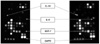
Chemiluminescent detection of the membranes for Human Inflammatory Cytokine/Receptor Gene Array demonstrated the expression of several genes. There was increased expression of some genes on the membrane of patients with rotator cuff disease compared with the controls. For example, IL-1A, IL-6 and SDF-1 were strongly expressed on the membrane of the rotator cuff disease patients, but there was no expression observed on the membrane from the control (Fig. 1).
3. cDNA Array Analysis for Inflammatory Cytokine/Receptor Gene Expression
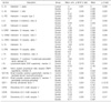
Most genes of the inflammatory cytokines and receptors analyzed with the array in the rotator cuff disease and control groups showed similar expression levels. However, there were significant differences in the expression of the genes from 20 cytokines between the two groups (Table 1).
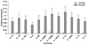
The expression of several interleukin genes (IL-1A, IL-1B, IL-6, IL-15, IL-16 and IL-17) was higher in patients with rotator cuff disease than in the controls (p<0.05) (Fig. 2). In addition, the expression of several interleukin receptor genes (IL-1R2, IL-6R, IL-12RB1, IL-12RB2, IL-13RA1 and IL-15RA) was higher in the patients with rotator cuff disease than the controls (p<0.05) (Fig. 2). The gene expression of IL-11, IL-12 and IL-13 was approximately twice that in the patient with rotator cuff disease than in the control. However, only one statistical analysis method revealed significant differences between the two groups.
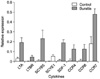
The expression of several inflammatory cytokine genes (LTA, SCYB5, SCYE1, and SDF-1) was higher in the patients with rotator cuff disease than the controls (p<0.05) (Fig. 3). Gene expression of lymphotoxin beta receptor (LTBR) and several chemokine (C-C motif) receptors (CCR4, CCR6 and CCR7) was higher in the patients with rotator cuff disease than the controls (p<0.05) (Fig. 3). The expression level of CCR 3 and tumor necrosis factor (TNF) was approximately twice that observed in the patients with rotator cuff disease than the control. However, only one statistical analysis method showed a significant difference in.
DISCUSSION
This study showed an increase in gene expression of several inflammatory cytokines and receptors in the subacromial bursa of patients with rotator cuff disease. While these findings improve the understanding of the complex biochemical processes that occur at the subacromial bursa in rotator cuff disease, some of these findings are consistent with previous studies. Gotoh et al.9) reported that IL-1 induced subacromial synovitis and there was a correlation between the level of IL-1 expression and the amount of shoulder pain, as assessed by the visual analog scale. They also suggested that the relationship between the expression of the IL-1B and IL-1 receptor antagonist in the subacromial bursa plays an important role in shoulder pain in rotator cuff disease. Szomor et al.28) reported that almost all subacromial bursa samples expressed IL-6 and IL-8 mRNA. On the other hand, there was no detectable IL-1B and TNF-α mRNA expression in a study of 17 patients with rotator cuff tears. Sakai demonstrated the increased expression of IL-1, TNF-α, TGF and bFGF in the subacromial bursa of patients with rotator cuff tear compared with that of anterior shoulder instability23). They suggested that these inflammatory cytokines and growth factors might play an important role in inflammation of the subacromial bursa.
In this study, the gene expression of many interleukins (IL-1A, IL-1B, IL-6, IL-15, IL-16 and IL-17) was higher at the subacromial bursa tissue in patients with rotator cuff disease than the controls. IL-1 is an inflammatory chemokine that recruits leukocytes for the host defense in an infection or inflammation2). IL-1 is also believed to induce pain through cyclooxygenase-2 (COX-2) expression, resulting in the production of prostaglandins (PGs). It was reported that IL-15 is produced by synoviocytes and synovial endothelial cells from RA patients, and stimulates the proliferation of T-lymphocytes16). IL-6 is released by a variety of cells including monocytes, macrophages, fibroblasts, and endothelial cells in response to inflammatory and profibrotic mediators such as IL-1B, TNF-α, platelet-derived growth factor (PDGF), and TGF-β29). The gene expression of cytokine receptors including IL-1R2), IL-6R, IL-12RB1, IL-12RB2, IL-13RA1 and IL15RA was higher in the patients with rotator cuff disease. IL-12 is a cytotoxic lymphocyte maturation factor and a natural killer cell stimulatory factor. IL-13 might be essential in regulating the inflammatory and immune responses17).
Chemokines, which are a short form of 'chemotactic cytokine', consist of 70-130 amino acids with four conserved cysteines12). The two main subfamilies, C-C and C-X-C chemokines are distinguished by the position of the first two cysteines, which are separated by a single amino acid (C-X-C) or adjacent (CC)12). In this study, the gene expressions of CCR4 (chemokine (C-C motif) receptor 4), CCR6 (chemokine (C-C motif) receptor 6) and CCR7 (chemokine (C-C motif) receptor 7) were increased significantly at the subacromial bursa of a patient with rotator cuff disease. All these genes are receptor genes for only the C-C chemokines subfamilies, not the C-X-C subfamilies. Although there is little data on the biological activity of these chemokines, particularly in the subacromial bursa, these results support the possible pathological role of these C-C subfamily chemokines and receptors in subacromial bursitis. In addition to the various cytokines identified, which play a role in inflammatory processes, this study also identified several novel chemokines that have not been previously considered. There were also significant increases in the gene expression of stromal cell derived factor-1 (SDF-1), small inducible cytokine subfamily E, member 1 (SCYE1, endothelial monocyte-activating) and small inducible cytokine superfamily B, member 5 (SCYB5, epithelial-derived neutrophil-activating) in the subacromial bursitis tissue. SDF-1 is a well known chemotactic factor in chronic inflammation of rheumatoid arthritis10,11,18,20,31). SDF-1 is a highly efficacious lymphocyte chemoattractant, and attracts lymphocytes and mononuclear cells from the blood stream into the site of inflammation4,5,18). Therefore, SDF-1 is a key cytokine that maintains the inflammatory condition. SCYE1 is a endotheial monocyte-activating polypeptide (EMAP) II, which is a proinflammatory cytokine originally isolated from the supernatants of methylcholanthrene (MethA)-induced fibrosarcoma cells14). However, there is little information on EMAP II expression in human tissue. EMAP II causes the chemotactic migration of polymorphonuclear leukocytes (PMNs) and monocytes3,13) and plays a role in antiangiogenesis3). SCYB5 is a epithelial-derived neutrophil-activating factor-78 (ENA-78), which is a 78 amino acid peptide with neutrophil-activating and chemotactic properties21). However, there is little data on ENA-78. One study reported that ENA-78 was produced by allograft tubule cells during human renal allograft rejection and caused leukocyte recruitment into the tubulointerstitial compartment26). It is believed that SDF-1, SCYE1 and SCYB5 play important roles in the induction of inflammation in bursal tissue.
The gene expression of lymphotoxin-α (LTA) and lymphotoxin-β receptor (LTBR) was higher in the rotator cuff disease group than in the control group (Table 1). LTA belongs to the TNF ligand family, which is a large number of structurally related proteins that play important roles in the immune and inflammatory response6). Lymphotoxin is expressed mainly in the lymphocyte compartment7). Its signaling through the lymphotoxin-β receptor is essential for inducing the key cytokines, chemokines and other factors that organize the intricate microenvironment within the lymphoid tissues and sustain chronic inflammation in autoimmune models ranging from diabetes to colitis1,7). These results suggest that LTA acts with LTBR in an inflammation reaction and plays a pathologic role in subacromial bursitis.
While these results represent novel findings regarding gene expression in the subacromial bursa of patients with rotator cuff disease, there were some limitations in this study. In some of the subacromial bursa tissue, the amount of RNA extracted was too small to analyze using a cDNA array technique. Since the subacromial bursa tissue was harvested during surgery, it is difficult to collect enough tissue for analysis. Therefore, the cDNA amplification was performed using the GEArray Ampolabeling-LPR kit Protocol. An adequate amount of cDNA with high quality for analysis could be obtained using this method. The cDNA Array technique with the GEAarry TMQ Series Kits is a new and powerful tool for screening a large number of genes simultaneously. However, there are some limitations of this method associated with the sensitivity12,30) and specificity12). King and Sinha pointed out several possible pitfalls that attenuated the power of a microarray, including the cellular heterogeneity of the tissue and comparability of various arrays15). With any array technique, additional confirmatory studies will be needed to corroborate the biological significance of the microarray findings. An attempt was made to address these issues by performing statistical analysis using two statistical tests; Mann Whitney U and Student's t test, which were analyzed by an experienced statistician. Continued investigations using polymerase chain reaction will be needed to further corroborate these findings. In this study, the control specimens of the subacromial bursa were acquired from patients with shoulder anterior instability and proximal humerus fractures. While the use of these bursa tissues may not represent a perfect normal control, several studies used patients with shoulder instability as the controls. Therefore, it is believed that this tissue was adequate9,23,32). All tissue specimens were harvested from the same location at the subacromial bursa tissue adjacent to the supraspinatus tendon insertion at the greater tuberosity.
CONCLUSION
There is a significant increase in the expression of many inflammatory cytokines in the subacromial bursa of a patient with rotator cuff disease. While further studies will be needed, these findings support a pathologic role of inflammatory cytokines in subacromial bursitis. These results are expected to provide the guidelines for further studies on the inflammation of subacromial bursa, and may ultimately lead to the design of a targeted treatment for subacromial bursitis without the side effects of the commonly used non-steroidal antiinflammatory agents and steroids.
 XML Download