

 PDF
PDF ePub
ePub Citation
 Print
Print


Abstract
Purpose
To evaluate the effectiveness, cosmetic and functional improvement of a supracondylar lateral closing wedge osteotomy of the humerus as a treatment for cubitus varus deformity in children.
Materials and Methods
Forty-eight children with cubitus varus underwent a lateral closing wedge osteotomy, and were followed up for at least 1 year.
Results
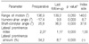
There were no complications such as a loss of correction, infection, or neurapraxia. The immediate postoperative lateral condylar prominence and secondary lazy S deformity was in proportion to the preoperative severity of the cubitus varus. However, it was lower at the last follow-up, and was related to the extent of preoperative cubitus varus, length of follow-up and age.
Figures and Tables
Fig. 1
(A) Lateral closing wedge osteotomy was performed. (B) Lazy 'S' deformity was observed on the radiograph of two months after the operation.
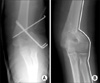
Fig. 2
(A) Humero-ulnar angle was measured on the anteroposterior radiograph of the elbow. (B) Shaft-condylar angle was measured on the lateral radiograph of the elbow.
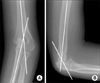
Fig. 3
(A) The angle of cubitus varus deformity was measured intraoperatively with C-arm fluoroscopy. (B) The operation was performed through a lateral approach. (C) We used two preset Kirschner's wires. (D) Osteotomized site was fixed with Steinmann pins. (E) Cubitus varus was corrected after surgery.
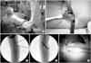
Fig. 4
(A) Lateral prominence index (LPI) is BC/AB. (B) Lateral prominence amount (LPA) is BC/AC × 100 (%).
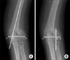
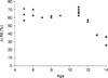
Fig. 5
(A) A six-year-old boy had the varus humeroulnar angle of 33° on the preoperative radiograph. (B) On the immediate postoperative radiograph, LPI was 3.23 and LPA was 45.3 percent. (C) On the radiograph after a 4-year-follow-up, the LPI was 1.01, and the LPA was zero percent.
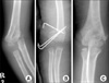
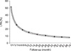
Fig. 7
The change in the amount of lateral prominence diminished with increasing age when the operation had been performed.
References
1. Barrett IR, Bellemore MC, Kwon YM. Cosmetic results of supracondylar osteotomy for correction of cubitus varus. J Pediatr Orthop. 1998. 18:445–447.

2. Bellemore MC, Barrett IR, Middleton RW, Scougall JS, Whiteway DW. Supracondylar osteotomy of the humerus for correction of cubitus varus. J Bone Joint Surg Br. 1984. 66:566–572.
3. Danielsson LG, Hussein S, el-Haddad I, Gupta RP. Staple fixation of osteotomy for cubitus varus. A simple technique used in II children. Acta Orthop Scand. 1991. 62:55–57.
4. Devnani AS. Lateral closing wedge supracondylar osteotomy of humerus for post-traumatic cubitus varus in children. Injury. 1997. 28:643–647.
5. Graham B, Tredwell SJ, Beauchamp RD, Bell HM. Supracondylar osteotomy of the humerus for correction of cubitus varus. J Pediatr Orthop. 1990. 10:228–231.
6. Griffin PP. Supracondylar fractures of the humerus. Pediatr Clin North Am. 1975. 22:477–486.
7. Høyer A. Treatment of supracondylar fracture of the humerus by skeletal traction in an abduction splint. J Bone Joint Surg Am. 1952. 24:623–637.
8. Hui JP, Torode IP, Chatterjee A. Medial approach for corrective osteotomy of cubitus varus: a cosmetic incision. J Pediatr Orthop. 2004. 24:477–481.
9. Ippolito E, Moneta MR, D'Arrigo C. Post-traumatic cubitus varus. Long-term follow-up of corrective supracondylar humeral osteotomy in children. J Bone Joint Surg Am. 1990. 72:757–765.
10. Karatosun V, Alekberov C, Alici E, Ardic CO, Aksu G. Treatment of cubitus varus using the Ilizarov technique of distraction osteogenesis. J Bone Joint Surg Br. 2000. 82:1030–1033.
11. King D, Secor C. Bow elbow(cubitus varus). J Bone Joint Surg Am. 1951. 33:572–576.
12. Kumar K, Sharma VK, Sharma R, Maffulli N. Correction of cubitus varus by French or dome osteotomy: A comparative study. J Trauma. 2000. 49:717–721.
13. LaBelle H, Bunnell WP, Duhaime M, Poitras B. Cubitus varus deformity following supracondylar osteotomy of the humerus in children. J Pediatr Orthop. 1982. 2:539–540.
14. Levin MJ, Horn BD, Pizzultillo PD. Treatment of posttraumatic cubitus varus in the pediatric population with humeral osteotomy and external fixation. J Pediatr Orthop. 1996. 16:597–601.
15. McCoy GF, Piggot J. Supracondylar osteotomy for cubitus varus. J Bone Joint Surg Br. 1988. 70:283–286.
16. Oppenheim WL, Clader TJ, Smith C, Bayer M. Supracondylar humeral osteotomy for traumatic childhood cubitus varus deformity. Clin Orthop Relat Res. 1984. 188:34–39.
17. Pankaj A, Dua A, Malhotra R, Bhan S. Dome osteotomy for posttraumatic cubitus varus: A surgical technique to avoid lateral condylar prominence. J Pediatr Orthop. 2006. 26:61–66.
18. Tachdjian MR. Smith AB, editor. Osteotomy for distal humerus for correction of cubitus varus. Pediatric orthopedics. 1972. Philadelphia: Saunders WB;1588–1591.
19. Tien YC, Chih HW, Lin GT, Lin SY. Dome corrective osteotomy for cubitus varus deformity. Clin Orthop Relat Res. 2000. 380:158–166.
20. Usui M, Ishii S, Miyano S, Narita H, Kura H. Three-dimensional corrective osteotomy for treatment of cubitus varus after supracondylar fracture of the humerus in children. J Shoulder Elbow Surg. 1995. 4:17–22.
21. Voss FR, Kasser JR, Trepman E, Simmons E, Hall JE. Uniplanar supracondylar humeral osteotomy with preset Kirschner wires for posttraumatic cubitus varus. J Pediatr Orthop. 1994. 14:471–478.
22. Williams PL, Warwick R. Gray's anatomy. 1980. 36th ed. Philadelphia: WB Saunders Co;365.
23. Wong HK, Lee EH, Balasubramaniam P. The lateral condylar prominence: a complication of supracondylar osteotomy for cubitus varus. J Bone Joint Surg Br. 1990. 72:859–861.
 XML Download