

 PDF
PDF ePub
ePub Citation
 Print
Print


An osteochondroma, which is also known as osteocartilaginous exostosis, is the most commonly occurring benign bone neoplasm, representing 42% to 50% of all bone tumors1,2,4,6). In one report of a series of 783 osteochondromas, only 15 osteochonromas were encountered in the tarsal region, and 10 of these were in the calcaneus10). An osteochondroma in the talus is very rare. We report the diagnosis and treatment of two cases of symptomatic osteochondroma in a rare location at the talus.
CASE REPORT
1. Case 1
A 36-year-old male presented with a painful mass on his right ankle. The patient first noticed a bean sized swelling on the dorsum of his right ankle 4 years earlier. The pain was exacerbated by sports activities. There was no history of trauma or other joint problems. The physical examination revealed a firm, non-mobile lump and tenderness over the anteromedial aspect of the ankle.
The range of motion of the right ankle was 5° in dorsiflexion. The dorsiflexion of his right ankle was less than the contralateral side, whereas the plantar flexion had a normal range of motion. The patient complained of pain upon forced dorsiflexion and plantar flexion.
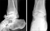
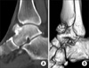
Radiography of the right ankle demonstrated a lobulated mass on the anteromedial part of the talus that was continuous with the cortex and medulla of the talus (Fig. 1). Computed tomography of the right ankle revealed a lobulated osseous mass arising from the talus, measuring 1.6×2.0×2.5 cm in size. The mass was connected to the medullary cavity of the talus (Fig. 2).
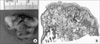
An excision was performed under general anesthesia. After a longitudinal medial incision, the mass was exposed and excised at the base of the stalk. The irregular surfaced mass consisted of a gray-white bony structure with a hard capsule. The pathology findings were consistent with an osteochondroma having the typical finding of a cartilage cap (Fig. 3). The patient was followed up for 20 months without recurrence. The range of motion of his right ankle was normal without pain.
2. Case 2
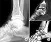
A 35-year-old male was referred to our hospital with a gradually progressing painless mass on the medial side of the left ankle. The patient had noticed a bean sized swelling 3 years ago. He felt intermittent pain while running or jogging, and a mechanical block in terms of a limitation of motion. He had no history of a previous injury to his left ankle. The physical examination revealed an egg sized hard lump that was palpated on dorsum of the talonavicular joint area without tenderness. There was neither tenderness nor acute inflammation. The dorsiflexion of the ankle was restricted to 0°, whereas the plantar flexion was normal (40°). The power of dorsiflexion and plantar flexion was normal. There was no neurovascular deficit. Radiography of the ankle revealed a 2×2 cm sized lobulated mass with a broad based stalk on the anteromedial part of the talus, which was connected with medullary cavity of the talus (Fig. 4).
An excisional biopsy was performed under general anesthesia. Using a medial skin incision, the mass was exposed and excised along with the periosteum at the base of the stalk. The irregular surfaced mass consisted of a gray-white bony structure with hard capsule. The pathology examination revealed an overlying cartilaginous cap that was consistent with an osteochondroma.
The patient was followed up for 15 months with no recurrence. The pain during exercise had subsided completely. The dorsiflexion was improved (20°) and the range of motion of his left ankle was normal.
DISCUSSION
An osteochondroma is the most common benign bone tumor and occurs most commonly in the proximal humerus, tibia, and distal femur8). Osteochondromas can occur in any bone that is preformed from cartilage. The most common locations are the long bones at the metaphyseal region8). It is rarely found in bones of the foot, and is even less common in the talus1,4,7,9). Osteochondromas are usually discovered during the first and second decades of life4). However, an osteochondroma in the talus is usually was discovered in the third to fifth decades1,4,7,9) including our cases.
An osteochondroma of the talus was first reported in 1984 by Fuselier et al4). They reported a solitary osteochondroma of the dorsum of the talus in a 22-year-old female presenting with ankle discomfort. They are found 2.0 cm long pedunculated osteochondroma, protruding from the dorsolateral head of the talus with multiple toe deformities.
In 1987, Chioros et al1) reported an atypical osteochondroma that originated from the posterior aspect of the talus in a 34-year-old male. In 2003, Erler et al2) reported a case of an osteochondroma located on the dorsum of the talus, which is similar to these cases, in 6-year-old boy without other foot deformities. There are a few other reports of osteochondroma in talus7,9).
A solitary osteochondroma is usually asymptomatic. However, an osteochondroma in the talus may represent with variable symptoms, including pain1), ankle swelling2), painless mass7,9), and a limited range of ankle motion1,4,7). This condition can be a spontaneous hemarthrosis of the ankle5).
Pain is usually caused by pressure and friction against the nerves and bones resulting in possible nerve irritation or a block of joint motion1). The mass can be in the form of an intraarticular loose body6). which is accompanied by severe painful limitation of motion. An osteochondroma can occur in the talar origin if it takes the form of an intraarticular loose body. Since an osteochondroma does not originate from the epiphysis of the ankle, it probably originates from the talus. In our cases, one complained of painful swelling with a limitation of ankle motion, and the other complained of painless swelling with a limitation of ankle motion.
Radiologically, an osteochondroma in the talus can be visualized as a protrusion from the host bone in a pedunculated2,4,9) or sessile1,7) manner as in the long bone. Both cases were the pedunculated types. Computed tomography of diagnosis of the medullary and cortical continuity between the lesion and talar bone is important for diagnosing an osteochondroma of the talus, as in the other solitary osteochondromas2,4).
The treatment of an asymptomatic osteochondroma of the talus might be just observation. However, surgical excision is a good treatment method for a symptomatic osteochondroma of the talus, as in our cases.
An osteochondroma is rarely found in the ankle but it should be included in a differential diagnosis with a painful or painless lump. An osteochondroma can detach from the stalk and can be found as an intraarticular loose body in an ankle joint. Unlike a simple bony protrusion, an extraperiosteal complete excision is the key to complete eradication and for preventing a recurrence.
 XML Download