

 PDF
PDF ePub
ePub Citation
 Print
Print


Synovial chondromatosis is an uncommon disorder of joints, bursae, and tendon sheaths. The etiology of the disease is unclear, but it involves the metaplasia of normal synovial cells to chondroblasts5). Synovial chondromatosis is characterized by the formation and growth of cartilaginous bodies that may then calcify or ossify within synovial tissue1). The usual treatment involves loose body removal and complete synovectomy.
Here, we describe a case of synovial chondromatosis of the fifth metacarpophalangeal (MP) joint.
CASE REPORT
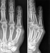
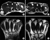
A 45-year-old, right-handed man presented with a 3-year history of pain, swelling and stiffness of the MP joint of the little finger of his right hand. He had no history of accident trauma or of a problem with other joints. Discomfort and progressive limitation of motion interfered with his daily activities. On examination, a non-tender, hard, and nodular mass about 2×2 cm in size was found and moderate tenderness along the joint line and a range of motion from 5° to 70° was evident, but sensation and perfusion in the little finger were normal. A plain X-ray showed diffuse calcification in the area of the swelling (Fig. 1), and magnetic resonance imaging (MRI) scan findings were consistent with synovial chondromatosis, which appeared to be localized to joint synovium and not to invade any surrounding structure (Fig. 2). We planned definitive surgery, i.e., loose body removal and complete synovectomy.
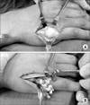
Under subscalenous regional anesthesia, with a tourniquet, surgical incision was performed through a longitudinal dorsal incision over the MP joint to expose the joint capsule. Division of the capsule revealed thickened inflammatory synovium, with numerous chondrous nodules in the capsule wall and lying free (Fig. 3A). These loose bodies were well contained within the dorsal pouch of synovium and capsule and between the joint surfaces. In edition the peripheral articular cartilage was eroded (Fig. 3B). Complete synovectomy was carried out and all loose bodies were removed.
Postoperatively, the patient was followed up for one year had a full active range of motion, with no MP joint instability.
DISCUSSION
Synovial chondromatosis is an uncommon condition characterized by the formation of multiple cartilaginous nodules in synovial tissue. It generally involves a large joint4), and its etiology has not been determined. The hand is rarely involved, but when it is, extra-articular structures, such as, the tenosynovium are usually affected8). Synovial chondromatosis tends to present between the ages of 20 and 50 and it is twice as common in men5). Its natural history is variable, and at presentation symptoms have usually been present from 3 months to 10 years. A correct diagnosis may not be made because of its rarity, especially if no calcification or ossification of cartilage is evident2). MRI is the best imaging modality as it shows the cartilaginous portion of the lesion in addition to any ossified areas3,7). Destruction of the joint space is rare, but if present, is confirmed at surgery. Our case had a positive radiologic finding, which included ossified loose bodies and periarticular erosion. However, the joint space was well preserved.
Treatment in most cases of intra-articular chondromatosis is by arthrotomy, loose body removal and synovectomy. This treatment is uniformly successful; all reported patients have experienced symptom resolution and an improved range of motion at follow-up.
Intra-articular synovial chondromatosis in the hand is a rare but benign condition, which should be considered in the differential diagnosis of patients presenting with a swollen, stiff or painful joint. The most effective treatment is surgical synovectomy.
 XML Download