

 PDF
PDF ePub
ePub Citation
 Print
Print


Abstract
Purpose
To evaluate the value of an open wedge high tibial osteotomy (HTO) using a Locking Compression Plate® (LCP®) as a surgical technique.
Materials and Methods
From May, 2003 to January, 2005, eleven open wedge HTO using LCP® were performed and the average follow-up period was 17.8 months. The knee score and function score for the clinical results, and the degree of varus deformity, the size of the joint space, the posterior tibial slope and the medial instability for radiography results were evaluated.
Results
The knee score improved from 54.8 points to 95.9 points, and the function score improved from 57.3 points to 88.2 points. The femorotibial angle was corrected from 4.1° varus to 9.9° valgus. The posterior tibial slope did not show any significant change. The size of the joint space increased from 3.3 mm to 4.3 mm. No medial instability was observed.
Figures and Tables
Fig. 1
Operative technique. (A) The pes anserinus was detached and the superficial MCL was detached from the poximal tibia (arrow: superficial MCL, arrow head: pes anserinus). (B) The increase in the posterior tibial slope was prevented by distracting the posterior site of the osteotomy.
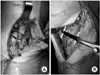
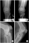
Fig. 2
A 43-year-old woman with left knee osteoarthritis. (A) Preoperative weight bearing anteroposterior view shows that the femorotibial angle is varus 4° and the size of the medial joint space was approximately 2 mm. (B) 2.5 years-postoperative weight bearing anteroposterior view shows that the femorotibial angle is valgus 11° and the medial joint space has increased by 4 mm. (C) Preoperative lateral view shows that the posterior tibial slope is 9°. (D) 2.5 year-postoperative lateral view shows that the posterior tibial slope is 8°.
References
1. Bae DK, Yoon KH, Kwon OS, Kim YC, Shin DJ. Results and survivorship of high tibial osteotomy. J Korean Orthop Assoc. 2002. 37:357–363.

2. Bauer GC, Insall J, Koshino T. Tibial osteotomy in gonarthrosis (osteo-arthritis of the knee). J Bone Joint Surg Am. 1969. 51:1545–1563.
3. Billings A, Scott DF, Camargo MP, Hofmann AA. High tibial osteotomy with a calibrated osteotomy guide, rigid internal fixation, and early motion. Long-term follow-up. J Bone Joint Surg Am. 2000. 82:70–79.
4. Brouwer RW, Bierma-Zeinstra SM, van Koeveringe AJ, Verhaar JA. Patellar height and the inclination of the tibial plateau after high tibial osteotomy. The open versus the closed-wedge technique. J Bone Joint Surg Br. 2005. 87:1227–1232.
5. Coventry MB, Bowman PW. Long-term results of upper tibial osteotomy for degenerative arthritis of the knee. Acta Orthop Belg. 1982. 48:139–156.
6. Coventry MB, Ilstrup DM, Wallrichs SL. Proximal tibial osteotomy. A critical long-term study of eighty-seven cases. J Bone Joint Surg Am. 1993. 75:196–201.
7. Fujisawa Y, Masuhara K, Shiomi S. The effect of high tibial osteotomy on osteoarthritis of the knee. An arthroscopic study of 54 knee joints. Orthop Clin North Am. 1979. 10:585–608.
8. Hernigou P, Medevielle D, Debeyre J, Goutallier D. Proximal tibial osteotomy for osteoarthritis with varus deformity. A ten to thirteen-year follow-up study. J Bone Joint Surg Am. 1987. 69:332–354.
9. Insall JN. Insall JN, Windsor RE, Scott WN, Kelly MA, Alietti P, editors. Osteotomy. Surgery of the knee. 1993. New York: Churchill Livingstone;635–676.
10. Insall JN, Joseph DM, Msika C. High tibial osteotomy for varus gonarthrosis. A long-term follow-up study. J Bone Joint Surg Am. 1984. 66:1040–1048.
11. Insall J, Salvati E. Patella position in the normal knee joint. Radiology. 1971. 101:101–104.
12. Katz MM, Hungerford DS, Krackow KA, Lennox DW. Results of total knee arthroplasty after failed proximal tibial osteotomy for osteoarthritis. J Bone Joint Surg Am. 1987. 69:225–233.
13. Kettelkamp DB, Leach RE, Nasca R. Pitfalls of proximal tibial osteotomy. Clin Orthop Relat Res. 1975. 106:232–241.
14. Koshino T, Murase T, Saito T. Medial opening-wedge high tibial osteotomy with use of porous hydroxyapatite to treat medial compartment osteoarthritis of the knee. J Bone Joint Surg Am. 2003. 85:78–85.
15. Kim SS, Lee HK, Lee SH, et al. A 5- to 13-year follow-up of high tibial osteotomy. J Korean Orthop Assoc. 1995. 30:494–501.
16. Lee JY, Seon JK, Song EK, Yoon TR, Cheon SY, Lim KY. Comparison of high tibial osteotomy: opening versus closing wedge osteotomy. J Korean Orthop Assoc. 2004. 39:790–796.
17. Lobenhoffer P, Simoni CD, Staubli AE. Open-wedge high-tibial osteotomy with rigid plate fixation. Techniques in Knee Surgery. 2002. 1:93–105.
18. Magyar G, Ahl TL, Vibe P, Toksvig-Larsen S, Lindstrand A. Open-wedge osteotomy by hemicallotasis or the closed-wedge technique for osteoarthritis of the knee. A randomised study of 50 operations. J Bone Joint Surg Br. 1999. 81:444–448.
19. Majima T, Yasuda K, Katsuragi R, Kaneda K. Progression of joint arthrosis 10 to 15 years after high tibial osteotomy. Clin Orthop Relat Res. 2000. 381:177–184.
20. Mankin HJ. The response of articular cartilage to mechanical injury. J Bone Joint Surg Am. 1982. 64:460–466.
21. Nakamura E, Mizuta H, Kudo S, Takagi K, Sakamoto K. Open-wedge osteotomy of the proximal tibia hemicallotasis. J Bone Joint Surg Br. 2001. 83:1111–1115.
22. Nicholas JA. The five-one reconstruction for anteromedial instability of the knee. Indications, technique, and the results in fifty-two patients. J Bone Joint Surg Am. 1973. 55:899–922.
23. Noyes FR, Goebel SX, West J. Opening wedge tibial osteotomy: the 3-triangle method to correct axial alignment and tibial slope. Am J Sports Med. 2005. 33:378–387.
24. Noyes FR, Mayfield W, Barber-Westin SD, Albright JC, HecHmann TP. Opening wedge high tibial osteotomy: an operative technique and rehabilitation program to decrease complications and promote early union and function. Am J Sports Med. 2006. 34:1262–1273.
25. Pace TB, Hofmann AA, Kane KR. Medial-opening high tibial osteotomy combined with Magnuson intraarticular debridement for traumatic gonarthrosis. J Orthop Tech. 1994. 2:21–28.
26. Song EK, Seol JY, Jeong KC, Choi J. Restoration of joint space of the knee after high tibial osteotomy. J Korean Knee Soc. 2002. 14:36–42.
27. Staubli AE, De Simoni C, Babst R, Lobenhoffer P. TomoFix: a new LCP-concept for open wedge osteotomy of the medial proximal tibia-early results in 92 cases. Injury. 2003. 34:Supple 2. B55–B62.
28. Sterett WI, Steadman JR. Chondral resurfacing and high tibial osteotomy in the varus knee. Am J Sports Med. 2004. 32:1243–1249.
29. Stoffel K, Stachowiak G, Kuster M. Open wedge high tibial osteotomy: biomechanical investigation of the modified Arthrex Osteotomy Plate (Puddu Plate) and the TomoFix Plate. Clin Biomech (Bristol, Avon). 2004. 19:944–950.
30. Sundaram NA, Hallett JP, Sullivan MF. Dome osteotomy of the tibia for osteoarthritis of the knee. J Bone Joint Surg Br. 1986. 68:782–786.
31. Tjornstrand BA, Egund N, Hagstedt BV. High tibial osteotomy: a seven-year clinical and radiographic follow-up. Clin Orthop Relat Res. 1981. 160:124–136.
32. Vainionpaa S, Laike E, Kirves P, Tiusanen P. Tibial osteotomy for osteoarthritis of the knee. A five to ten-year follow-up study. J Bone Joint Surg Am. 1981. 63:938–945.
33. Windsor RE, Insall JN, Vince KG. Technical considerations of total knee arthroplasty after proximal tibial osteotomy. J Bone Joint Surg Am. 1988. 70:547–555.
34. Yasuda K, Majima T, Tsuchida T, Kaneda K. A 10- to 15-year follow-up observation of high tibial osteotomy in medial compartment osteoarthrosis. Clin Orthop Relat Res. 1992. 282:186–195.
35. Weale AE, Lee AS, MacEachern AG. High tibial osteotomy using a dynamic axial external fixator. Clin Orthop Relat Res. 2001. 382:154–167.
36. Wright JM, Heavrin B, Begg M, Sakyrd G, Sterett W. Observations on patellar height following opening wedge proximal tibial osteotomy. Am J Knee Surg. 2001. 14:163–173.
 XML Download