

 PDF
PDF ePub
ePub Citation
 Print
Print


A lumbosacral deformity is a fixed spinal deformity that cannot be corrected with traction, suspension, or side bending. Surgery is often recommended because it is difficult to control it with a brace but the method of correcting the deformity is controversial. A trapezoidal wedge resection osteotomy of L5 is generally performed for the treatment of congenital scoliosis due to a sacral malformation. We introduce the surgical technique and show that a wedge resection lumbar osteotomy can correct a coronal and sagittal deformity.
CASE REPORT
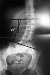
A 41-year-old female complained of a 25-year history of lower back pain and buttock pain accompanied with intermittent radiating pain to the left leg. The coronal imbalance was 3.8 cm, as determined by the distance between the C7 plumb line and the center sacral line. The scoliosis angle using the Cobb method was 22° (Fig. 1). The sacral malformation was a fixed lumbosacral deformity that could not be corrected by traction or forced side bending. A trapezoidal wedge resection osteotomy of the L5 body was planned for the correction of rigid scoliosis.
With the spine exposed posteriorly, the paraspinal muscles were stripped subperiosteally from the spinous processes to the tips of the transverse processes at the L3 to S2 levels. The posterior arch, spinous process, laminae and articular processes of L5 was removed and the dura mater and nerve roots of L5 and S1 on both sides were verified. The pedicles of L5 were removed using a rongeur and the cancellous bone was partially removed through the site of the pedicles by curettage.
The transverse processes were also morcellized with an electrical burr and the normal soft tissues were retracted as far lateral as possible. Pedicular screws were inserted bilaterally into the L3, L4, S1, and S2 levels under fluoroscopic guidance and a temporary rod was inserted at the left side. After meticulous homeostasis had been obtained with bipolar electrocauterization, the dura mater and nerve roots were retracted carefully, and the posterior wall and side walls of the L5 vertebral body were removed.
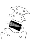
The lengths of the resection at the pedicle level were 22 mm on the right side and 9 mm on the left, which were the same as determined in preoperative planning (Fig. 2). A intravertebral trapezoidal wedge resection was performed gradually toward the anterior vertebral body in a so called piecemeal fashion using dissectors, curettes and pituitary forceps The anterior wall was also removed using a Kerrison rongeur, taking particular care to leave the soft tissue anterior to the body intact. This was followed by confirming that the canal was clear of any residual compression.
The temporary rod was removed and the precontoured rods were inserted one by one and the osteotomy was closed in a compression manner. During closure, the nerve roots and dura mater were observed under direct vision of the avoidance of compression. Posterior fusion was performed in a meticulous manner, decorticating all posterior elements and applying autogenous corticocancellous bone at all levels instrumented. Before closing the wound, radiographs of the spine were obtained to determine the coronal and sagittal balance.
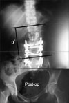
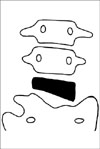
Postoperatively, the scoliosis angle was corrected to 0° and the coronal imbalance had disappeared (Fig. 3 and 4). The patient was allowed to walk 2 weeks after surgery. The well-molded thoracolumbosacral orthosis was applied for 4 months when solid fusion was apparent on the radiographs. At the 2 year follow-up, the patient returned to normal physical activity without any deformity or low back pain.
DISCUSSION
A posterior closing-wedge vertebral osteotomy has been proposed as corrective surgery for kyphosis8). The neurological and vascular complications of this technique are much lower than the Smith-Petersen type anterior opening-wedge osteotomy, producing a sharp lordotic angle and an elongation of the anterior column. However, most early studies reported the clinical results only in ankylosing spondylitis with kyphotic deformity5,8,9).
Since the year 2000, a transpedicular wedge resection osteotomy, or so called pedicle subtraction osteotomy, have been used to treat various fixed sagittal imbalances, including iatrogenic kyphosis, post-traumatic kyphosis, degenerative kyphosis, post-infectious kyphosis, idiopathic scoliosis and tumors1,3,4).
Congenital scoliosis is most often observed in the thoracic area and least in the lumbosacral area10). Only limited cases of coronal corrections in the treatment of congenital scoliosis have been reported1,6,7). Our case was a congenital scoliosis due to a sacral malformation, which was considered to be a unilateral failure of the vertebral formation of S1.
A spinal deformity at the lumbosacral region poses a unique problem. The lack of a mobile spine below the anomaly often results in early truncal decompensation and a long compensatory curve above, which can progresses with time. A lumbosacral deformity frequently produces functional impairment and neurological involvement. Pain is a common symptom in later years.
There are only a few reports on a vertebral column resection for the treatment of rigid coronal decompensation. Most were accomplished using combined anterior and posterior approaches in one or two stages2). Vertebral column resections through the posterior approach only were reported in the treatment of congenital scoliosis and kyphoscoliosis. However, the cases reported were hemivertebrae at the thoracic or lumbar level7).
We performed a modification of a vertebral wedge osteotomy in a patient with a rare condition, and confirmed the correction capacity of this procedure, both clinically and radiologically. The characteristics of this vertebral osteotomy were a trapezoidal shaped resection in the coronal plane and a three-column resection in the sagittal plane. It is believed that an intravertebral trapezoidal wedge resection osteotomy is an effective technique for correcting a fixed coronal and a sagittal deformity in various spinal deformities.
 XML Download