

 PDF
PDF ePub
ePub Citation
 Print
Print


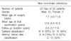
Abstract
Purpose
To evaluate the clinical and radiological results of a new innominate osteotomy in Legg-Calve-Perthes' disease (LCPD).
Materials and Methods
This study examined 25 hips that were treated with a new innominate osteotomy for LCPD. The treatment involved the anterior half of the ilium being osteomized in a direction of 45° to the coronal plane and 30° to 45° to the sagittal plane, and the posterior half of the ilium being cut using a Gigli saw according to the conventional method. The mean follow-up duration was 5.5 years. Stable interposition of the bone block was achieved using a single biodegradable screw in 8 hips, and without any fixation device in 17 hips.
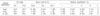
Results
The clinical results according to the criteria of Robinson were good in 20 hips. Twelve hips was graded as good by the Mose method, according to the criteria of Stulberg, 8 hips were included in class I, 6 hips in class II, 8 hips in class III, and 3 hips in class IV. The mean center-edge angle improved from 19.4° to 30.2°.
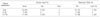
Figures and Tables
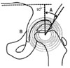 | Fig. 1The center-edge angle (A) was measured from this line drawn from center of the ossific nucleus to the lateral ossified margin of the acetabulum and a line perpendicular to a third line joining the inferior margins of the sacroiliac joints. The template of mose (B) with concentric rings 2 mm apart are superimposed on the femoral head on the AP roentenogram, and the Mose method was used to assess the distance between the inner and outer best fitting ring superimposed on the femur head.
|
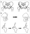 | Fig. 2(A) Diagram of the original Salter osteotomy. (B) This photographs show the site and direction of the original slater innominate osteotomy.
|
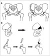 | Fig. 3(A) Diagram of our modification. (B) This photographs showing the site and direction of the modification. Maximum bony contact between the triangular fragment and host bone can be achieved.
|
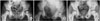 | Fig. 4(A) Preoperative pelvis AP radiograph in a 6-year-old-girl with Legg-Calve-Perthes' disease of the Left hip (Caterall group IV). (B) This is the pelvis AP radiograph taken at 3 month after the new Salter innominate osteotomy. (C) A radiograph taken at the final examination when the patient was 11 years of age shows a Stulberg class II and a good Mose method.
|
References
1. Canale ST, D'Anca AF, Cotler JM, Snedden HE. Innominate osteotomy in Legg-Calve-Perthes disease. J Bone Joint Surg Am. 1972. 54:25–40.
2. Catterall A. The natural history of Perthes' disease. J Bone Joint Surg Br. 1971. 53:37–53.

3. Herring JA, Neustadt JB, Williams JJ, Early JS, Browne RH. The lateral pillar classification of Legg-Calve-Perthes disease. J Pediatr Orthop. 1992. 12:143–150.
4. Ingman AM, Paterson DC, Sutherland AD. A comparison between innominate osteotomy and hip spica in the treatment of Legg-Perthes' disease. Clin Orthop Relat Res. 1982. 163:141–147.
5. Ishida A, Kuwajima SS, Laredo Filho J, Milani C. Salter innominate osteotomy in the treatment of severe Legg-Calve-Perthes disease: clinical and radiographic results in 32 patients (37 hips) at skeletal maturity. J Pediatr Orthop. 2004. 24:257–264.
6. Kitakoji T, Hattori T, Kitoh H, Katoh M, Ishiguro N. Which is a better method for Perthes' disease: femoral varus or Salter osteotomy? Clin Orthop Relat Res. 2005. 430:163–170.
7. Millis MB, Hall JE. Transiliac lengthening of the lower extremity. A modified innominate osteotomy for the treatment of postural imbalance. J Bone Joint Surg Am. 1979. 61:1182–1194.
8. Moberg A, Hansson G, Kaniklides C. Results after femoral and innominate osteotomy in Legg-Calve-Perthes disease. Clin Orthop Relat Res. 1997. 334:257–264.
9. Mose K. Methods of measuring in Legg-Calve-Perthes disease with special regard to the prognosis. Clin Orthop Relat Res. 1980. 150:103–109.
10. Olney BW, Asker MA. Combined innominate and femoral osteotomy for the treatment of severe Legg-Calve-Perthes disease. J Pediar Orthop. 1985. 5:645–651.
11. Robinson HJ Jr, Putter H, Sigmond MB, O'Connor S, Murray KR. Innominate osteotomy in Perthes disease. J Pediatr Orthop. 1988. 8:426–435.
12. Salter RB. Experimental and clinical aspects of Perthes' disease. J Bone Joint Surg Br. 1966. 48:393.
13. Salter RB. The present status of surgical treatment for Legg-Calve-Perthes disease. J Bone Joint Surg Am. 1984. 66:961–966.
14. Salter RB. Treatment by innominate osteotomy. AAOS Instruct Course Lect. 1973. 22:309–316.
15. Sponseller PD, Desai SS, Millis MB. Comparison of femoral and innominate osteotomies for the treatment of Legg-Calve-Perthes disease. J Bone Joint Surg Am. 1988. 70:1131–1139.
16. Stulberg SD, Cooperman DR, Wallensten R. The natural history of Legg-Calve-Perthes disease. J Bone Joint Surg Am. 1981. 63:1095–1108.
17. Vukasinovic Z, Slavkovic S, Milickovic S, Siqeca A. Combined salter innominate osteotomy with femoral shortening versus other methods of treatment for Legg-Calve-Perthes disease. J Pediar Orthop B. 2000. 9:28–33.
18. Yoon TR, Rowe SM, Chung JY, Song EK, Mulyadi D, Anwar IB. A new innominate osteotomy in Perthes' disease. J Pediatr Orthop. 2003. 23:363–367.
19. Wiberg G. Studies on dysplastic acetabula and congenital subluxation of the hip joint with special refernce to the complication of osteoarthritis. Acta Chir Scand. 1993. 83:Suppl 58. S1–S135.
 XML Download