

 PDF
PDF ePub
ePub Citation
 Print
Print


Abstract
Purpose
To compare the results of two different surgical methods (translation vs rod derotation) in a correction of double thoracic adolescent idiopathic scoliosis (AIS) using pedicle screw instrumentation.
Materials and Methods
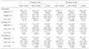
Forty-seven patients with double thoracic AIS treated by pedicle screw instrumentation were reviewed retrospectively after a minimum follow-up of 2 years. The patients were divided into two groups; TR group (translation method, n=14) and RD group (rod derotation, n=33). There were no significant differences in the preoperative curve characteristics between the two groups.
Results
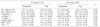
In the TR group, the preoperative upper thoracic curve of 37±4° improved to 24±4° (35% correction), and the lower thoracic curve of 52±9° improved to 18±5° (65% correction). In the RD group, the preoperative upper thoracic curve of 40±7° improved to 19±7° (51% correction), and the lower thoracic curve of 56±12° was improved to 16±6° (72% correction). The correction of the upper and lower thoracic curves was significantly better in the RD group (p<0.05). Thoracic sagittal kyphosis was corrected from 21° to 24° in the TR group and from 18° to 26° in the RD group. There was no significant difference in the spinal balance, shoulder height difference, T1 tilt and fusion extent. The operating time and the amount of blood loss was 231 minutes and 2050ml in the TR group and 263 minutes and 3217ml in the RD group, respectively (p<0.05).
References
1. Chang KW. Cantilever bending technique for treatment of large and rigid scoliosis. Spine. 2003. 28:2452–2458.

2. Cil A, Pekmezci M, Yazici M, et al. The validity of Lenke criteria for defining structural proximal thoracic curves in patients with adolescent idiopathic scoliosis. Spine. 2005. 30:2550–2555.
3. Cotrel Y, Dubousset J, Guillaumat M. New universal instrumentation in spinal surgery. Clin Orthop Relat Res. 1988. 277:10–23.
4. Delorme S, Labelle H, Aubin CE, et al. A three-dimensional radiographic comparison of Cotrel-Doubousset and Colorado instrumentation for the correction of idiopathic scoliosis. Spine. 2000. 25:205–210.
5. Delorme S, Labelle H, Aubin CE, et al. Intaoperative comparison of two instrumentation techniques for the correction of adolescent idiopathic scoliosis. Rod rotation and translation. Spine. 1999. 24:2011–2017.
6. Gardner-Morse M, Stokes IA. Three-dimensional simulations of the scoliosis derotation maneuver with Cotrel-Dubousset instrumentation. J Biomech. 1994. 27:177–187.
7. Goshi K, Boachie-Adjei O, Moore C, Nishiyama M. Thoracic scoliosis fusion in adolescent and adult idiopathic scoliosis using posterior translational corrective techniques (Isola): is maximum correction of the thoracic curve detrimental to the unfused lumbar curve? Spine J. 2004. 4:192–201.
8. Kim DS, Suk SI, Kim WJ, Rhee HC. Comparison of apical Z-axis derotation between rod derotation (RD) and vertebrae to rod (VTR) methods in idiopathic thoracic scoliosis. J Korean Soc Spine Surg. 2000. 7:253–258.
9. King HA, Moe JH, Bradford DS, Winter RB. The selection of fusion levels in thoracic idiopathic scoliosis. J Bone Joint Surg Am. 1983. 65:1302–1313.
10. Labelle H, Dansereau J, Bellefleur C, et al. Comparison between preoperative and postoperative three-dimensional reconstructions of idiopathic scoliosis with the Cotrel-Dubousset procedure. Spine. 1995. 20:2487–2492.
11. Lee CK, Denis F, Winter RB, Lonstein JE. Analysis of the upper thoracic curve in surgically treated idiopathic scoliosis. A new concept of the double thoracic curve pattern. Spine. 1993. 18:1599–1608.
12. Lee DH, Lee JH, Kim SH, et al. Tactics for surgical treatment of the double thoracic scoliosis: significance of T1 tilt, first rib elevation and correction ratio. J Korean Soc Spine Surg. 2002. 9:106–114.
13. Lenke LG, Bridwell KH, O'Brien MF, Baldus C, Blanke K. Recognition and treatment of the proximal thoracic curve in adolescent idiopathic scoliosis treated with Cotrel-Dubousset instrumentation. Spine. 1994. 19:1589–1597.
14. Muschik M, Schlenzka D, Robinson PN, Kupferschmidt C. Dorsal instrumentation for idiopathic adolescent thoracic scoliosis: rod rotation versus translation. Eur Spine J. 1999. 8:93–99.
15. Steib JP, Dumas R, Mitton D, Skalli W. Surgical correction of scoliosis by in situ contouring: a detorsion analysis. Spine. 2004. 29:193–199.
16. Suk SI, Kim WJ, Lee CS, et al. Indications of proximal thoracic curve fusion in thoracic adolescent idiopathic scoliosis: recognition and treatment of double thoracic curve pattern in adolescent idiopathic scoliosis treated with segmental instrumentation. Spine. 2000. 25:2342–2349.
17. Suk SI, Lee CK, Kim WJ, Chung YJ, Park YB. Segmental pedicle screw fixation in the treatment of thoracic idiopathic scoliosis. Spine. 1995. 20:1399–1405.
18. Webb JK, Burwell RG, Cole AA, Lieberman I. Posterior instrumentation in scoliosis. Eur Spine J. 1995. 4:2–5.
19. Winter RB. The idiopathic double thoracic curve pattern. Its recognition and surgical management. Spine. 1989. 14:1287–1292.
 XML Download