

 PDF
PDF ePub
ePub Citation
 Print
Print


Abstract
Purpose
This study reviewed the outcome of the surgical treatment of osteofibrous dysplasia (OFD) involving the long bones in order to demonstrate the necessity of an extraperiosteal excision.
Materials and Methods
This study was a retrospective review of 10 cases of OFD, who underwent surgical treatment from August 1996 to August 2003. All cases were diagnosed by the final histology. There were 5 males and 5 females with a mean age of 12.4 years (range, 4.2-42.6 years). Seven, 1 and 2 involved the tibia, fibular, and femur, respectively. For surgical treatment, an extraperiosteal excision, subperiosteal excision or curettage and bone grafting was selected. Recurrences and bony union were observed using the serial radiographs. The mean follow-up duration was 4.2 years (range, 2.2-6.6 years).
Results
Six, 1 and 3 patients underwent an extraperiosteal excision, subperiosteal excision, and curettage and bone grafting, respectively. Two patientswho underwent curettage and one patient who underwent subperiosteal excision suffered recurrences. There were two cases of non-union in the extraperiosteal excision group which required bone grafting. There were no infections or pathological fractures.
Figures and Tables
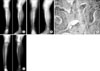 | Fig. 1A case of localized extraperiosteal excision. (A) 4.2-year old boy who suffered from a fracture twice visited showing a growing mass in the lower leg. An eccentric intracortical osteolytic lesion with a sclerotic margin was demonstrated at the proximal third of the tibia. (B) A localized extraperiosteal excision with a fibular strut bone graft was performed because of the sudden expansion of the mass, which was confined to less than 50% of the bony circumference. (C) Irregularly arranged bony trabeculae lying within a relatively hypocellular fibroblastic proliferation is the typical finding of osteofibrous dysplasia. (D) Two years and 8 months after surgery, the radiograph shows the evidence of union and no finding of recurrence.
|
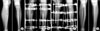 | Fig. 2A case of segmental extraperiosteal excision. (A) A 7.3-year old boy suffered lower leg pain and a growing mass. A eccentric osteolytic lesion with a sclerotic margin was demonstrated. (B) A segmental extraperiosteal excision and external fixation with Ilizarov was performed because of the extensive lesion over 50% of the bony circumference, which resulted in bony weakness. (C) Bone transport was performed for the bony defect. (D) Four years and 2 months after surgery, the radiograph shows evidence of union and no recurrence.
|
References
1. Benassi MS, Campanacci L, Gamberi G, Ferrari C, Picci P, Sangiorgi L, Campanacci M. Cytokeratin expression and distribution in adamantinoma of the long bones and osteofibrous dysplasia of tibia and fibula: an immunohistochemical study correlated to histogenesis. Histopathology. 1994. 25:71–76.

2. Cambell CJ, Hawk T. A variant of fibrous dysplasia, osteofibrous dysplasia. J Bone Joint Surg Am. 1982. 64:231–236.
3. Campanacci M, Laus M. Osteofibrous dysplasia of the tibia and fibular. J Bone Joint Surg Am. 1981. 63:367–375.
4. Georgen TG, Dickman PS, Resnick D, Saltzstein SL, O'Dell CW, Akeson WH. Long bone ossifying fibroma. Cancer. 1977. 39:2067–2072.
5. Hahn SB, Chun IM, Shin KH. Treatment of ossifying fibroma. J Korean Orthop Assoc. 1995. 30:1759–1766.
6. Hahn SB, Kang ES, Jahang JS, Park BM, Choi JC. Ossifying fibroma. J Korean Orthop Assoc. 1990. 25:606–613.
7. Hazelbag HM, Taminiau AH, Fleuren GJ, Hogenboorn PCW. Adamantinoma of the long bones. A clinicopathological study of thirty-two patients with emphasis on histological subtype, precursor lesion, and biological behavior. J Bone Joint Surg Am. 1994. 76:1482–1499.
8. Kahn LB. Adamantinoma, osteofibrous dysplasia and differenciated adamantinoma. Skeletal Radiol. 2003. 32:245–258.
9. Kempson RL. Ossifying fibroma of the long bone: A light and electron microscopic study. Arch Pathol. 1966. 88:218–233.
10. Lee HK. Skeletal oncology. 1996. 1st ed. Seoul: Choi-Shin Medical Book;314–315.
11. Lee RS, Weitzel S, Eastwood DM, Monsell F, Pringle J, Cannon SR, Briggs TW. Osteofibrous dyslplasia of the tibia is there a need for a radical surgical approach? J Bone Joint Surg Br. 2006. 88:658–664.
12. Lee SH, Suh SW, Hong JS, Wang JH, Rho YJ. Osteofibrous dysplasia of the tibia in children. J Korean Orthop Assoc. 2001. 36:601–606.
13. Markel SF. Ossifying fibroma of lone bone. Its distinction from fibrous dysplasia and its association with adamantinoma of long bone. Am J Clin Pathol. 1978. 69:91–97.
14. Mirra JM. Bone tumors, clinical, radiologic, and pathologic correlations. 1998. 1st ed. Philadelphia: Lea & Febiger;1217–1229.
15. Nakashima Y, Yamamuro T, Fujiwara Y, Kotoura Y, Mori E, Hamashima Y. Osteofibrous dysplasia. A study of 12 cases. Cancer. 1983. 52:909–914.
16. Ozaki T, Hamada M, Sugihara S, Kunisada T, Mitani S, Inoue H. Treatment outcome of osteofibrous dysplasia. J Pediatr Orthop, Part B. 1998. 7:199–202.
17. Springfield DS, Rosenberg AE, Mankin HJ, Mindell ER. Relationship between osteofibrous dysplasia and adamantinoma. Clin Orthop Relat Res. 1994. 309:234–244.
18. Wang JW, Shih CH, Chen WJ. Osteofibrous dysplasia, ossifying fibroma of long bones, A report of four cases and review of the literature. Clin Orthop Relat Res. 1992. 278:235–243.
 XML Download