

 PDF
PDF ePub
ePub Citation
 Print
Print


Abstract
Purpose
To evaluate the diagnosis and treatment of the carpal tunnel syndrome (CTS) due to space occupying lesion (SOL)s.
Materials and Methods
14 patients (15 cases) that underwent surgery from 1992 to 2002 for CTS due to SOL were studied. The average age was 51 years. There were 6 men and 8 women. Mean follow up period was 16 months. In patients with swelling or tenderness on the area of wrist flexion creases, MRI and/or CT scan were additionally taken as well as the carpal tunnel view. We performed conventional open transverse carpal ligament release and removal of SOL.
Results
The types of lesion were, in three cases tuberculosis tenosynovitis, nonspecific tenosynovitis in three cases, gout in one case, mass in four cases, and abnormal palmaris longus hypertrophy in one case. Bony lesions were, in one case Kienböck's disease (stage III), neglected volar dislocation of lunate in two cases. Following surgery, all cases showed alleviation of symptoms.
Figures and Tables
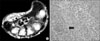 | Fig. 1(A) MRI shows hypertrophied flexor digitorum profundus tenosynovium in carpal tunnel (Black arrow: hypertrophied tenosynovium). (B) Pathologic findings were compatible with tuberculosis tenosynovitis. There were caseous necrosis and granuloma with Langerhan's giant cell (black arrow) and lymphocytic infiltration (H-E stain, ×200).
|
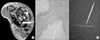 | Fig. 2(A) MRI shows tophi infiltration between flexor digitorum profundus tenosynovium and carpal bones (white arrow: tophi infiltration). (B) Urate crystal and lymphocyte infiltration show chronic tophaceous arthritis (H-E stain, ×200). (C) When examined with a polarizing filter, negative birefringence was noted.
|
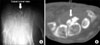
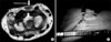 | Fig. 3(A) MRI shows hypertrophied palmaris longus is compressing median nerve in carpal tunnel (white arrow: hypertrophied palmaris longus muscle). (B) Open transverse carpal ligament release and hypertrophied palmaris longus excision was performed.
|
References
1. Backhouse KM, Churchill-Davidson D. Anomalous palmaris longus muscle producing carpal tunnel-like compression. Hand. 1992. 7:22–24.

2. Bagatur AE, Zorer G. The carpal tunnel syndrome is a bilateral disorder. J Bone Joint Surg Br. 2001. 83:655–658.
3. Bechtol CO. Grip test; the use of a dynamometer with adjustable handle spacings. J Bone Joint Surg Am. 1954. 36:820–824.
4. Coessens B, De Mey A, Lacotte B, Vandenbroeck D. Carpal tunnel syndrome due to an haemangioma of the median nerve in a 12-year-old child. Ann Chir Main Memb Super. 1991. 10:255–257.
5. Cseuz KA, Thomas JE, Lambert EH, Love JG, Lispocomb PR. Long-term results of operation for carpal tunnel syndrome. Mayo Clin Proc. 1966. 41:232–241.
6. Edwards AJ, Sill BJ, Macfarlane I. Carpal tunnel syndrome due to dystrophic calcification. Aust N Z J Surg. 1984. 54:491–492.
7. Evangelisti S, Reale VF. Fibroma of tendon sheath as a cause of carpal tunnel syndrome. J Hand Surg Am. 1992. 17:1026–1027.
8. Chen WS. Median-nerve neuropathy associated with chronic anterior dislocation of the lunate. J Bone Joint Surg Am. 1995. 77:1853–1857.
9. Horch RE, Allmann KH, Laubenberger J, Langer M, Stark GB. Median nerve compression can be detected by magnetic resonance imaging of the carpal tunnel. Neurosurgery. 1997. 41:76–83.
10. Kamolz LP, Schrogendorfer KF, Rab M, Girch W, Gruber H, Frey M. The precision of ultrasound imaging and its relevance for carpal tunnel syndrome. Surg Radiol Anat. 2001. 23:117–121.
11. Kang HJ, Park SY, Shin SJ, Kang ES, Hahn SB. Tuberculous tenosynovitis presenting as carpal tunnel syndrome -two cases-. J Korean Soc Surg Hand. 2000. 5:137–141.
12. Kerrigan JJ, Bertoni JM, Jaeger SH. Ganglion cysts and carpal tunnel syndrome. J Hand Surg Am. 1988. 13:763–765.
13. Kremchek TE, Kremchek EJ. Carpal tunnel syndrome caused by flexor tendon sheath lipoma. Orthop Rev. 1988. 17:1083–1085.
14. Lee KS, Woo KJ, Shim JH, Lee GH. The clinical study of grip and pinch strength in normal Korean adult. J Korean Orthop Assoc. 1995. 30:1589–1597.
15. Nakamichi K, Tachibana S. Ultrasonography in the diagnosis of carpal tunnel syndrome caused by an occult ganglion. J Hand Surg Br. 1993. 18:174–175.
16. Nakamichi K, Tachibana S. Unilateral carpal tunnel syndrome and space-occupying lesions. J Hand Surg Br. 1993. 18:748–749.
17. Pai CH, Tseng CH. Acute carpal tunnel syndrome caused by tophaceous gout. J Hand Surg Am. 1993. 18:667–669.
18. Weiss APC, Steichen JB. Synovial sarcoma causing carpal tunnel syndrome. J Hand Surg Am. 1992. 17:1024–1025.
19. Yesildag A, Kutluhan S, Sengul N, et al. The role of ultrasonographic measurements of the median nerve in the diagnosis of carpal tunnel syndrome. Clin Radiol. 2004. 59:910–915.
 XML Download