

 PDF
PDF ePub
ePub Citation
 Print
Print


Abstract
Purpose
This study examined the results of treatment according to the stage and size of a spontaneous osteonecrosis lesion of the knee (SONK) in the patients over 60 years of age.
Materials and Methods
Twenty-two knees from 19 patients over 60 years of age were treated for spontaneous osteonecrosis of the knee at our institution between January, 2000, and June, 2004. The mean follow time was 20.7 months. The condylar ratios, proportion of the lesion size to the condyle were measured. The size was obtained by multiplying the values from the anteroposterior and lateral radiographs. The stages were classified radiographically, and the treatment results were analyzed according to the size and stage. Conservative treatment was performed if the ratio and size were <40% and 5.0 cm2, respectively. Arthroplasty was performed if the ratio or size was >40% or 5.0 cm2. A paired T-test, Spearman correlation test and Wilcoxon test were used for the statistical evaluation.
Results
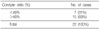
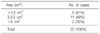
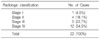
There was a higher prevalence in females (15 patients, 79%), and the mean age was 65 years (46-77 years). Bilateral involvement was observed in 3 patients. The lesions involved mainly the medial femoral condyle (21 cases, 95%). Seven cases (31%) had a condylar ratio <40% and 15 cases (69%) had a condylar ratio >40%. Conservative treatments were performed in 5 cases staged radiographically as I or II and there were no significant changes in the knee scores (ρ=0.931). Of the 17 cases staged III and IV radiographically, conservative treatment and arthroplasty were performed in 7 and 10 cases, respectively. Seven cases, in whom conservative treatments had been performed, showed a decreased in the knee scores compared with the increased knee scores in 10 patients treated with arthroplasty (ρ=0.943).
Conclusion
An accurate diagnosis and measurement of the size and staging of spontaneous osteonecrosis of knee in patients over 60 years of age is important for proper treatment. Clinically, no further progression of symptoms is visible on grade I and II spontaneous osteonecrosis of the knee after conservative treatment. However, arthroplasty improves the clinical results in patients with radiological grade III and IV osteonecrosis compared with conservative treatment.
Figures and Tables
Fig. 1
(A) Simple radiography for measuring the size of the lesion on the anterior-posterior view. The condylar ratio and transverse diameter can be measured. (B) Simple radiography for measuring the size of the lesion on the lateral view. The depth can also be measured. *a: Maximum transverse width of the medial femoral condyle on the anteriorposterior radiography, b: Transverse width of the lesion on anteriorposterior radiography, c: The greatest length of the lesion on lateral radiography.
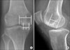
Fig. 2
Outcome of conservative treatment in stages I and II. There is a strong correlation in the outcome of conservative treatment (ρ=0.931). *AKSS: American Knee Society Score.
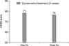
Fig. 3
(A) On the initial simple radiography we cannot identify the lesion in the medial condyle of the femur (stage I). (B) After conservative treatment (12 months), the follow-up radiography showed a collapse of the medial femoral condyle (arrow).
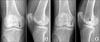
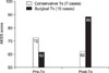
Fig. 4
Outcome of conservative and surgical treatment in stages III and IV. There is a statistical difference between the outcomes of conservative treatment and surgical treatment (p<0.005). *AKSS: American Knee Society Score

References
1. Aglietti P, Insall JN, Buzzi R, Deschamps G. Idiopathic osteonecrosis of the knee. Aetiology, prognosis and treatment. J Bone Joint Surg Br. 1983. 65:588–597.

2. Ahlbäck S, Bauer GC, Bohne WH. Spontaneous osteonecrosis of the knee. Arthritis Rheum. 1968. 11:705–733.
3. Akgun I, Kesmezacar H, Ogut T, Kebudi A, Kanberoglu K. Arthroscopic microfracture treatment for osteonecrosis of the knee. Arthroscopy. 2005. 21:834–843.
4. Forst J, Forst R, Heller KD, Adam G. Spontaneous osteonecrosis of the femoral condyle: causal treatment by early core decompression. Arch Orthop Trauma Surg. 1998. 117:18–22.
5. Havel PE, Ebraheim NA, Jackson WT. Steroid-induced bilateral avascular necrosis of the lateral femoral condyles. A case report. Clin Orthop Relat Res. 1989. 243:166–168.
6. Lecouvet FE, van de Berg BC, Maldague BE, et al. Early irreversible osteonecrosis versus transient lesions of the femoral condyles: prognostic value of subchondral bone and marrow changes on MR imaging. Am J Roentgenol. 1998. 170:71–77.
7. Lotke PA, Abend JA, Ecker ML. The treatment of osteonecrosis of the medial femoral condyle. Clin Orthop Relat Res. 1982. 171:109–116.
8. Lotke PA, Ecker ML, Alavi A. Painful knees in older patients: radionuclide diagnosis of possible osteonecrosis with spontaneous resolution. J Bone Joint Surg Am. 1977. 59:617–621.
9. Mont MA, Baumgarten KM, Rifai A, Bluemke DA, Jones LC, Hungerford DS. Atraumatic osteonecrosis of the knee. J Bone Joint Surg Am. 2000. 82:1279–1290.
10. Mont MA, Tomek IM, Hungerford DS. Core decompression for avascular necrosis of the distal femur: long term followup. Clin Orthop Relat Res. 1997. 334:124–130.
11. Motohashi M, Morii T, Koshino T. Clinical course and roentgenographic changes of osteonecrosis in the femoral condyle under conservative treatment. Clin Orthop Relat Res. 1991. 266:156–161.
12. Muheim G, Bohne WH. Prognosis in spontaneous osteonecrosis of the knee. Investigation by radionuclide scintimetry and radiography. J Bone Joint Surg Br. 1970. 52:605–612.
13. Rozing PM, Insall J, Bohne WH. Spontaneous osteonecrosis of the knee. J Bone Joint Surg Am. 1980. 62:2–7.
14. Soucacos PN, Johnson EO, Soultanis K, Vekris MD, Theodorou SJ, Beris AE. Diagnosis and management of the osteonecrotic triad of the knee. Orthop Clin North Am. 2004. 35:371–381.
15. Soucacos PN, Xenakis TH, Beris AE, Soucacos PK, Georgoulis A. Idiopathic osteonecrosis of the medial femoral condyle. Classification and treatment. Clin Orthop Relat Res. 1997. 341:82–89.
16. Weiner ES, Abeles M. Aseptic necrosis and glucocorticosteroids in systemic lupus erythematosus: a reevaluation. J Rheumatol. 1989. 16:604–608.
17. Yamamoto T, Bullough PG. Spontaneous osteonecrosis of the knee: the result of subchondral insufficiency fracture. J Bone Joint Surg Am. 2000. 82:858–866.
 XML Download