

 PDF
PDF ePub
ePub Citation
 Print
Print


Abstract
Purpose
To examine the correlation between the Navigation and radiographic measurements for the postoperative mechanical axis and component position in total knee arthroplasty.
Materials and Methods
From December 2005 to May 2006, Navigation assisted MIS TKA was performed on 46 knees of 34 patients. After fixing the components, the mechanical axis (MA) of the lower extremity, femoral component position to the mechanical axis in the coronal plane (θ), tibial component position in the coronal (β) and sagittal (δ) planes was measured using the Navigation. Two observers measured the same angles using the postoperative follow-up radiographs. The measurements were compared and the correlation between the Navigation and radiographic measurement was analyzed.
Results
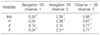
The average Navigation measurements were valgus 0.02±1.09° for MA, varus 0.05±0.96° for θ, valgus 0.02±0.86° for β and 4.03±1.25° for δ. The mean radiographic measurements of observer 1 were valgus 0.71±3.73° for MA, valgus 1.14±1.72° for θ, valgus 0.90±1.47° for β and 4.51±2.03° for δ. Those of observer 2 were valgus 0.12±3.39°, valgus 0.16±1.96°, valgus 0.30±1.65° and 3.85±1.60°, respectively.
Conclusion
The average of measurement for the component position showed a statistically significant difference in the MA (p=0.001), θ (p=0.000) and β (p=0.000) among three groups. There were a relatively high correlation between observer 1 and 2 for the radiographic measurements (r=0.67-0.96). However there was a very low correlation between the Navigation and radiographic measurements (r=0.10-0.39). Therefore, the possibility of a difference between the Navigation and radiographic measurement need to be considered when evaluating the component position.
Figures and Tables
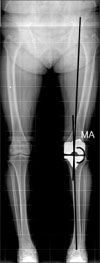
Fig. 2
The θ angle is the coronal femoral component angle against the femoral mechanical axis. The β angle is the coronal tibial component angle against the tibial mechanical axis.
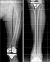
Fig. 3
The δ angle was formed between the mechanical axis of the tibia and the line of the tibial plate in the sagittal plane (posterior slope).
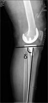
Table 1
Measured Angle of the Component and Mechanical Axis (mean±SD, degree)

MA, Mechanical axis; θ, Femoral component angle between the femoral mechanical axis and the femoral component; β, Tibial component angle between the tibial mechanical axis and the tibial component (+: valgus, -: varus); δ, Posterior slope of tibial component against sagittal tibial mechanical axis (+: posterior slope, -: anterior slope).
References
1. Anderson KC, Buehler KC, Markel DC. Computer assisted navigation in total knee arthroplasty: comparison with conventional methods. J Arthroplasty. 2005. 20:7 Suppl 3. 132–138.
2. Bach CM, Steingruber IE, Peer S, Nogler M, Wimmer C, Ogon M. Radiographic assessment in total knee arthroplasty. Clin Orthop Relat Res. 2001. 385:144–150.

3. Bae DK, Yoon KH, Song SJ, Kim SG, Park KJ. Intraoperative versus postoperative measurement in total knee arthroplasty using computer-assisted orthopaedic surgery (CAOS): accuracy of CAOS. J Korean Orthop Assoc. 2005. 40:168–173.
4. Bolognesi M, Hofmann A. Computer navigation versus standard instrumentation for TKA: a single-surgeon experience. Clin Orthop Relat Res. 2005. 440:162–169.
5. Choi HR, Lee SS, Park JS, Lee BI. Comparative analysis for radiographic measurement of component position between conventional and navigation assisted total knee arthroplasty. J Korean Knee Soc. 2006. 18:32–38.
6. Ewald FC. The knee society total knee arthroplasty roentgenographic evaluation and scoring system. Clin Orthop Relat Res. 1989. 248:9–12.
7. Haaker RG, Stockheim M, Kamp M, Proff G, Breitenfelder J, Ottersbach A. Computer-assisted navigation increases precision of component placement in total knee arthroplasty. Clin Orthop Relat Res. 2005. 433:152–159.
8. Kim SJ, MacDonald M, Hernandez J, Wixson RL. Computer assisted navigation in total knee arthroplasty: improved coronal alignment. J Arthroplasty. 2005. 20:7 Suppl 3. 123–131.
9. Mont MA, Fairbank AC, Yammamoto V, Krackow KA, Hungerford DS. Radiographic characterization of aseptically loosened cementless total knee replacement. Clin Orthop Relat Res. 1995. 321:73–78.
10. Seon JK, Song EK. The accuracy of lower extremity alignment in a total knee arthroplasty using computer-assisted navigation system. J Korean Orthop Assoc. 2004. 39:566–571.
11. Sparmann M, Wolke B, Czupalla H, Banzer D, Zink A. Positioning of total knee arthroplasty with and without navigation support. A prospective, randomized study. J Bone Joint Surg Br. 2003. 85:830–835.
12. Stulberg SD. How accurate is current TKR instrumentation? Clin Orthop Relat Res. 2003. 416:177–184.
13. Stulberg SD, Loan P, Sarin V. Computer-assisted navigation in total knee replacement: results of an initial experience in thirty-five patients. J Bone Joint Surg Am. 2002. 84:Suppl 2. 90–98.
14. Swanson KE, Stocks GW, Warren PD, Hazel MR, Janssen HF. Does axial limb rotation affect the alignment measurements in deformed limbs? Clin Orthop Relat Res. 2000. 371:246–252.
15. Tew M, Waugh W. Tibiofemoral alignment and the results of knee replacement. J Bone Joint Surg Br. 1985. 67:551–556.
 XML Download