Abstract
Purpose
Isoflavones are rich in soybean and are known to affect bone formation. This study examined the effects and modes of action of isoflavones on the differentiation of C2C12 myoblasts in the presence of the bone morphogenetic protein (BMP)-4.
Materials and Methods
The isoflavones, daidzein, genistein or equol, and/or BMP-4 were added alone or in combination to C2C12 myoblasts. After 72 hours culture, the cells were stained for the early osteoblastic differentiation marker, alkaline phosphatase (ALP). The ALP activity was determined by comparing the color of the stained images as well as by spectrophotometry. The expression profiles of the extracellular matrix (ECM) genes responsible for the extensive remodeling at the cell surface were analyzed using agene expression microarray after treating thesamples with daidzein.
Results
ALP staining of BMP-4 or the isoflavones-treated cells showed that BMP-4 increased the activity of ALP in a dose dependent manner, whereas the isoflavones alone did not induced any remarkable increase. However, the ALP activity increased when the cells were treated with BMP-4 and any of the three isoflavones. The macrogen mouse MAC array data showed that the ECM genes, Mmp13 and Mmp3, were up-regulated by daidzein, whereas Col4a2, Col5a1 and Mmp9 were down-regulated.
The role of estrogens and estrogen-like molecules, including isoflavones, in regulating the bone cell activities is essential for understanding the etiology and treatment of post-menopausal osteoporosis. Isoflavonoids, which are found abundant in soybeans, are a group of phytochemicals that show affinity for the estrogen receptor11,22). Isoflavonoids have been shown to be effective in preventing bone loss in ovariectomized rats and postmenopausal women17). Moreover, genistein was found to inhibit the bone loss caused by an estrogen deficiency in mice, and an analogue of coumestrol has been reported to increase the activity of ALP (an osteoblastic phenotype marker) in osteoblast-like cells9). These results suggest that isoflavones prevent bone loss and promote bone formation. In addition, it has been reported that isoflavones participate in the apoptosis and differentiations of several cell types6,8,24). Unlike estrogen, the consumption of isoflavones has been suggested to reduce the risk of breast and prostate cancer12).
Phytoestrogens have the potential to maintain bone health and delay or prevent osteoporosis, and several studies have examined the dose-dependent effects on bone and their modes of action4,14,23). Some reports concluded that the mechanism of action underlying the prevention of bone loss by isoflavones appears to differ from that of estrogen and the selective estrogen receptor modulators5,15). Whilst bothestrogen and raloxifene prevent bone loss by reducing bone resorption, the isoflavones are antiresorptive only at chronic high doses. Therefore, they have been reported to either inhibit or not affect the bone turnover rate. Moreover, neither estrogen nor raloxifene blocks the elevated bone resorption rates induced by an ovariectomy or menopause5). On the other hand, certain isoflavones, including genistein, have been shown to modestly stimulate the osteoblast characteristics, such as, the synthesis of total proteins and mineralized matrix deposition in immature and mature osteoblast cell cultures26). Hence, while isoflavone treatment may prevent bone loss, in part, by modulating the balance between bone formation and resorption, the mechanism underlying the enhanced bone formation after an isoflavone treatment in vivo is unknown.
C2C12 cells were derived from mouse myoblasts, and have the potential to differentiate into osteoblasts. Therefore, they are composed of committed osteoblastic precursor cells3). Many studies have examined the conversion of C2C12 cells into an osteoblast lineage when mediated by BMP2,10,27). It was hypothesized that C2C12 cells might provide useful information about the effects of isoflavones and BMP-4 on osteoblast differentiation. This study examined the in vitro effects of daidzein, genistein, or equol and/or BMP using C2C12 cells to determine the bioactivity of isoflavones in the presence or absence of BMP-4 on the bone metabolism.
The low passage mouse pluripotential mesenchymal precursor line, C2C12, was purchased from American Type Culture Collection (Manassas, VA). BMP-4 was obtained from R & D Systems (Minneapolis, MN) and was prepared as a stock solution (1 mg/ml) and stored at 4℃. The isoflavones (daidzein, genistein and Equol) was purchased from PKC Biochemicals (Woburn, MA) and was reconstituted in dimethyl sulfoxide (DMSO) at a 1000× stock concentration of 5 and 50 mM, respectively. The solutions were stored at -20℃. The cells were maintained in Dulbecco's modified Eagle medium (DMEM, Irvine Scientific, Santa Ana, CA) containing 20% fetal bovine serum (FBS, Invitrogen, Carlsbad, CA), supplemented with 50 units/ml penicillin and 50 µg/ml streptomycin (Mediatech Mediatech, Herndon, VA).
For the differentiation assays, the C2C12 cells were plated at an initial density of ca. 5×10⁴ cells/cm2. BMF-4 at 1, 3, or 10 ng/ml (final concentration) was added alone or in combination with 5 or 50 µM (final concentration) of either daidzein, genistein, or equol in 2% DMEM medium. The medium was renewed after 3 days.
The substrate used for the ALP assays was purchased from Sigma Chemical (St. Louis, MO). The ALP activity was visualized as follows. The medium was aspirated from the C2C12 cells cultured in 12 well plates, and the cells were then rinsed with phosphate-buffered saline (PBS), formaldehyde fixed for 10 min, and incubated in 300 µl of a Western blue stabilized substrate for ALP. The reaction was quenched after 30 min by removing the reaction buffer and washing once with PBS. The ALP activity was determined by comparing the colors of the scanned images.
The ALP activity was quantified using a previously described protocol. The medium was aspirated from the osteoblasts cultured in 12 well plates. The cells were rinsed with PBS and sonicated for 3×10 secs in cold lysis buffer (50 mM Tris (pH 7.5), 0.1% Triton X-100). The ALP activity in the cleared supernatant was assayed using reverse transcriptase (RT) in a reaction buffer containing 0.1 M 2 amino-2 methyl-1 propanol and 2 mM MgCl2 at pH 10.5, for 30 min using p-nitrophenyl phosphate (Sigma Chemical, St. Louis, MO). The reactions were quenched by adding 0.1 M NaOH, and the activities were determined by measuring the light absorbance at 410 nm and comparing results with standard p-nitrophenol solutions and the appropriate blank. The results are expressed as micromoles of p-nitrophenol produced per min per mg of protein. The ALP level was normalized to the protein concentration using Bicinchoninic Acid (BCA) Protein Assay Reagent (Pierce Chemical, Rockford, IL) that was determined spectrophotometrically at 562 nm and compared with the standard protein solutions.
The total RNA was isolated from a 10 cm template after 96 hours of culture with daidzein. RNAeasy kits (QIAGEN, Valencia, CA) were used, and the RNA concentrations were quantified by measuring the absorbance at 260 nm. The labeled probes were prepared by reverse transcription of 5 µg of RNA in the presence of dATP (PerkinElmer, Wellesley, MA). The probes were hybridized to Illumina arrays (Sentrix mouse Ref-8 Expression Bead Chips, Illumina, Inc., San Diego, CA) overnight. After thorough washing according to the manufacturer's protocol, the raw data was obtained. The scans were performed using an Applied Biosystems 1700 chemiluminescent Microarray Analyzer version 1.1.0. The differences in themicroarray intensities were normalized and grouped using Avadis Prophetic 3.3 version (Strand Genomics Pvt. Ltd., USA) by Quartile normalization, the 2-fold change method, and K-means cluster (Euclidean method/Complete linkage). The data was then entered into a database and analyzed graphically.
Daidzein dose-dependently induced C2C12 osteoblast differentiation in the presence of various BMP-4 concentrations. The C2C12 cell ALP activity was significantly higher after treatment with 50 µM daidzein than after 5 µM daidzein, as evidenced by the significantly higher level of ALP staining. In contrast to the cells treated with daidzein plus BMP-4, the cells treated with daidzein alone showed little color development (Fig. 1).
Treatment with genistein plus BMP-4 exhibited mild ALP activity at 5 µM genistein but there was significant induction at 50 µM. C2C12 cells treated with genistein alone showed only slight color changes.
The addition of 5 µM to 50 µM equol dose-dependently enhanced the ALP staining intensity compared with BMP-4 alone. As was observed with the other two isoflavones, equol alone was ineffective in inducing C2C12 differentiation, as evidenced by the weak blue staining.
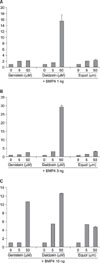
In order to quantify the ALP activity induced by isoflavones, the C2C12 myoblasts were treated separately with the isoflavones or with BMP-4. As shown in Fig. 2 (A-C), 50 uM of daidzein, equol or genistein synergistically induced ALP activity at BMP-4 concentrations of 1-10 ng/ml. In particular, daidzein appeared to induce ALP activity efficiently at the lower BMP-4 concentrations (i.e., 1 to 3 ng/ml)(Fig. 2).
This array technique was used to examine the RNA samples from the C2C12 cells treated with daidzein. The genes associated with the ECM glycoproteins, e.g., Leprel1, Nid1, Efemp1 and Ltbp3, were down-regulated, whereas Mfap3 and Fbln1 were up-regulated. The genes associated with the ECM linker proteins, e.g., Lamc1, Lama2, Lama5 were down-regulated. The genes associated with the ECM structural proteins, e.g., Col5a1, Col4a2, Col1a2 and Col8a1 were also down-regulated (Fig. 3). Among the other ECM associated genes, e.g., Mmp13 and Mmp3 were up-regulated but Mmp9 and Pcolce2 were down-regulated.
Osteogenin and the related BMPs are members of the TGF-β supergene family that are involved in a number of physiological events including embryonic development and skeletal tissue maintenance and repair20). In vitro, BMPs induce the differentiation of various types of cells to the osteoblastic lineage. These cells include undifferentiated mesenchymal cells, bone marrow stromal cells, and preosteoblasts. Moreover, BMP-2, 4, 6, 7, and 9 have been reported to significantly induce ALP activity in preosteoblastic C2C12 cells10).
An association between the ectoenzyme ALP and matrix vesicles during osteoblast differentiation and mineralization is required for ester phosphate hydrolysis at the sites of mineralization in order to provide ionic phosphate for calcium-phosphate formation1). Previous studies, using the ALP inhibitor levamisole, have reported the importance of ALP in the commitment of bone preosteoblasts to mineralization13). Indeed, it has been suggested that intracellular ALP participates in the regulation of cellular differentiation19). In the present study, ALP was used as a marker of osteoblastic differentiation of C2C12 cells. The results showed that the ALP activity increased appreciably by a treatment with the isoflavones plus BMP-4 whereas the isoflavones administered alone had no significant effect on osteoblastic differentiation. This suggests that isoflavones play a role in bone metabolism through growth factors such as BMP-4. Isoflavones are known weak agonist for estrogen receptors, while estrogen acts as an antiresorptive agent by inhibiting osteoclasts rather than by acting as a strong promoter of osteoblast production. However, the presence of isoflavones becomes meaningful when and where there are growth factors like BMPs. The action of isoflavones on bone development is expected to be more apparent in settings where the production of endogenous estrogens and/or BMPs are decreasing, such as in ovariectomized mice and/or heterozygotes of estrogen/BMPs.
Considerable attention has been given to the expression of specific gene products in osteoblasts16,25) but the comprehensive profiles of the changes in intracellular gene expression during differentiation to the osteoblast phenotype were not accessible before the introduction of gene array technology. Previous studies have shown that cells can be induced to differentiate to osteoblasts under a variety of conditions. In this study, gene array technology was used to chart the expressional changes in hundreds of gene during the osteoblastic differentiation of C2C12 mouse myoblasts. Gene expression was analyzed using a mouse gene array panel, which included a large number of genes encoding the structural ECM proteins. The results showed that the expression of many genes was altered in a seemingly unstructured manner. However, the up-regulation of Mmp13 and the down-regulation of Col4a2, Col5a1 and Mmp9 might bemeaningful in terms of osteoblastic differentiation. Along with MMP-14, MMP-9 (92-kDa gelatinase; gelatinase B) and MMP-13 (collagenase-3) have been reported to be the most abundant proteinases that regulate cellular migration, changes in the extracellular matrix and apoptosis in growth plate cartilage17). On the other hand, Mmp2 knock-out (-/-) mice have been reported to exhibit opposing bone phenotypes due to an impaired osteocytic canalicular networkand decreased bone mineral density in the limb and trunk bones but increased bone volume in the calvariae7). Type IV collagens including Col4a1 are the major components of the basement membrane in blood vessels, and are expressed ubiquitously in the basement membranes during the early developmental stages. Type V collagen (Col5), in addition to types I and XXIV collagens, is known to be a component of mineralized bone and corneal, whereas types II, XI, and XXVII collagen are the components of cartilage21). Although these MMPs and collagens are widely perceived to be essential structural and functional components during bone and cartilage development, the contribution of individual gene expression is unclear. Therefore, studies using knock-out mice with single or multiple genes deleted will be needed. Overall, these findings suggest that daidzein may affect osteoblastic differentiation in part by regulating the expression of the ECM associated proteins at the transcription level. However, further study will be needed to determine the mechanism whereby isoflavones affects osteoblast differentiation.
In summary, results of this study provide evidence that isoflavones weakly induce the expression of the osteoblastic marker, ALP, in C2C12 cells (a pre-osteoblastic cell line), particularly in the presence of BMP-4. This suggests that isoflavones may act with growth factors such as BMP-4 during the bone metabolism. Mouse illumina expression chip arrays showed that the expression of a subset of ECM associated genes are modulated by daidzein but the significance of these changes in the osteogenic process has yet to be determined. Overall these results suggest that isoflavones cause expressional changes in the ECM associated proteins that are known to play key roles in the osteogenic process, at the gene level. Comparative animal studies on bone development, bone density, and extracellular bone mineralization in mice fed daidzein with/without BMP should provide a deeper understanding of the effects of isoflavones on bone homeostasis.
Figures and Tables
References
2. Chandra A, Itakura T, Yang Z, et al. Neurogenesin-1 differentially inhibits the osteoblastic differentiation by bone morphogenetic proteins in C2C12 cells. Biochem Biophys Res Commun. 2006. 344:786–791.

3. Cheng H, Jiang W, Phillips FM, et al. Osteogenic activity of the fourteen types of human bone morphogenetic proteins (BMPs). J Bone Joint Surg Am. 2003. 85:1544–1552.

4. Dang ZC, Lowik C. Dose-dependent effects of phytoestrogens on bone. Trends Endocrinol Metab. 2005. 16:207–213.

5. Fanti P, Monier-Faugere MC, Geng Z, et al. The phytoestrogen genistein reduces bone loss in short-term ovariectomized rats. Osteoporos Int. 1998. 8:274–281.

6. Gu Y, Zhu CF, Iwamoto H, Chen JS. Genistein inhibits invasive potential of human hepatocellular carcinoma by altering cell cycle, apoptosis, and angiogenesis. World J Gastroenterol. 2005. 11:6512–6517.

7. Inoue K, Mikuni-Takagaki Y, Oikawa K, et al. A crucial role for matrix metalloproteinase-2 in osteocytic canalicular formation and bone metabolism. J Biol Chem. 2006. 281:33814–33824.
8. Ito C, Murata T, Itoigawa M, et al. Induction of apoptosis by isoflavonoids from the leaves of Millettia taiwaniana in human leukemia HL-60 cells. Planta Med. 2006. 72:424–429.
9. Kanno S, Hirano S, Kayama F. Effects of phytoestrogens and environmental estrogens on osteoblastic differentiation in MC3T3-E1 cells. Toxicology. 2004. 196:137–145.

10. Katagiri T, Yamaguchi A, Komaki M, et al. Bone morphogenetic protein-2 converts the differentiation pathway of C2C12 myoblasts into the osteoblast lineage. J Cell Biol. 1994. 127(6 Pt 1):1755–1766.
11. Kuiper GG, Lemmen JG, Carlsson B, et al. Interaction of estrogenic chemicals and phytoestrogens with estrogen receptor beta. Endocrinology. 1998. 139:4252–4263.
12. Kim H, Peterson TG, Barnes S. Mechanisms of action of the soy isoflavone genistein: emerging role for its effects via transforming growth factor beta signaling pathways. Am J Clin Nutr. 1998. 68:Suppl 6. S1418–S1425.

13. Klein BY, Gal I, Segal D. Studies of the levamisole inhibitory effect on rat stromal-cell commitment to mineralization. J Cell Biochem. 1993. 53:114–121.

14. Knight DC, Eden JA. A review of the clinical effects of phytoestrogens. Obstet Gynecol. 1996. 87(5 Pt 2):897–904.
15. Krishnan V, Heath H, Bryant HU. Mechanism of action of estrogens and selective estrogen receptor modulators. Vitam Horm. 2000. 60:123–147.

16. Lee KS, Kim HJ, Li QL, et al. Runx2 is a common target of transforming growth factor beta1 and bone morphogenetic protein 2, and cooperation between Runx2 and Smad5 induces osteoblast-specific gene expression in the pluripotent mesenchymal precursor cell line C2C12. Mol Cell Biol. 2000. 20:8783–8792.
17. Malemud CJ. Matrix metalloproteinases: role in skeletal development and growth plate disorders. Front Biosci. 2006. 11:1702–1715.

18. Morabito N, Crisafulli A, Vergara C, et al. Effects of genistein and hormone-replacement therapy on bone loss in early postmenopausal women: a randomized double-blind placebo-controlled study. J Bone Miner Res. 2002. 17:1904–1912.

19. Narisawa S, Hofmann MC, Ziomek CA, Millán JL. Embryonic alkaline phosphatase is expressed at M-phase in the spermatogenic lineage of the mouse. Development. 1992. 116:159–165.

20. Ripamonti U, Reddi AH. Growth and morphogenetic factors in bone induction: role of osteogenin and related bone morphogenetic proteins in craniofacial and periodontal bone repair. Crit Rev Oral Biol Med. 1992. 3:1–14.

21. Segev F, Héon E, Cole WG, et al. Structural abnormalities of the cornea and lid resulting from collagen V mutations. Invest Ophthalmol Vis Sci. 2006. 47:565–573.

22. Verma SP, Goldin BR. Effect of soy-derived isoflavonoids on the induced growth of MCF-7 cells by estrogenic environmental chemicals. Nutr Cancer. 1998. 30:232–239.

23. Wang CF, Kurzer MS. Effects of phytoestrogens on DNA synthesis in MCF-7 cells in the presence of estradiol or growth factors. Nutr Cancer. 1998. 31:90–100.

24. Wu HJ, Chan WH. Genistein protects methylglyoxal-induced oxidative DNA damage and cell injury in human mononuclear cells. Toxicol In Vitro. 2007. 21:335–342.

25. Xiao ZS, Hinson TK, Quarles LD. Cbfa1 isoform overexpression upregulates osteocalcin gene expression in non-osteoblastic and pre-osteoblastic cells. J Cell Biochem. 1999. 74:596–605.

26. Yamaguchi M, Sugimoto E. Stimulatory effect of genistein and daidzein on protein synthesis in osteoblastic MC3T3-E1 cells: activation of aminoacyl-tRNA synthetase. Mol Cell Biochem. 2000. 214:97–102.


 PDF
PDF ePub
ePub Citation
 Print
Print


 XML Download