

 PDF
PDF ePub
ePub Citation
 Print
Print


Abstract
Purpose
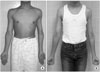
To examine the clinical results of a corrective dome osteotomy for a cubitus varus and valgus deformity.
Materials and Methods
Between January 1998 and April 2005, nineteen patients with a cubitus varus or valgus deformity were treated with a corrective dome osteotomy. The mean age of the patients was 29.5 years and the mean follow-up period was 39 months (range, 15 to 95 months). A dome osteotomy was performed along the circle centered approximately 1 cm distally from the olecrenon tip. Internal fixation was performed with multiple K-wires or plates.
Results
Bony union was achieved in 18 cases. In the cubitus varus group, the carrying angle was corrected from a mean varus of 17.9° to a mean valgus of 5.9°. The lateral prominence angle (LPI) was corrected from a mean of 15.6% to a mean of -7.6%. In the cubitus valgus group, the carrying angle was corrected from a mean valgus of 36° to 6.7°. The LPI was corrected from a mean -31% to -1.3%. On the functional assessment, 12, 5 and 2 cases showed excellent, good and fair outcomes, respectively.
Figures and Tables
Fig. 1
The lateral prominence index (LPI) was calculated using the dollowing formula: (AB-BC)/AC×100, where B is the crosslink between a line connecting the lateral prominence, A, the medial prominence, C, and the longitudinal mid-humeral axis.
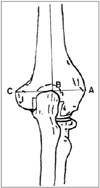
Fig. 2
Dome osteotomy was performed along the circle with a radius of approximately 3 cm centered about 1 cm distally from the olecrenon tip. The black circle indicates the center of rotation.
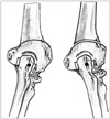
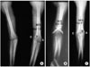
Fig. 4
(A) A 9-year old male had a cubitus varus deformity (carrying angle: varus 13°). (B) Dome osteotomy and fixation with K-wires were performed. (C) The carrying angle was corrected to valgus 5°.
References
1. Arnold JA, Nasca RJ, Nelson CL. Supracondylar fractures of the humerus: the role of dynamic factors in prevention of deformity. J Bone Joint Surg Am. 1977. 59:589–595.
2. Bellemore MC, Barrett IR, Middleton RW, Scougall JS, Whiteway DW. Supracondylar osteotomy of the humerus for correction of cubitus varus. J Bone Joint Surg Br. 1984. 66:566–572.

3. Chess DG, Leahey JL, Hyndman JC. Cubitus varus: significant factors. J Pediatr Orthop. 1994. 14:190–192.
4. DeRosa GP, Graziano GP. A new osteotomy for cubitus varus. Clin Orthop Relat Res. 1988. 236:160–165.
5. French PR. Varus deformity of the elbow following supracondylar fractures of the humerus in children. Lancet. 1959. 2:439–441.
6. Ippolito E, Moneta MR, D'Arrigo C. Post-traumatic cubitus varus. Long-term follow-up of corrective supracondylar humeral osteotomy in children. J Bone Joint Surg Am. 1990. 72:757–765.
7. Labelle H, Bunnell WP, Duhaime M, Poitras B. Cubitus varus deformity following supracondylar fractures of the humerus in children. J Pediatr Orthop. 1982. 2:539–546.
8. Laupattarakasem W, Mahaisavariya B, Kowsuwon W, Saengnipanthkul S. Pentalateral osteotomy for cubitus varus. Clinical experiences of a new technique. J Bone Joint Surg Br. 1989. 71:667–670.
9. Oppenheim WL, Clader TJ, Smith C, Bayer M. Supracondylar humeral osteotomy for traumatic childhood cubitus varus deformity. Clin Orthop Relat Res. 1984. 188:34–39.
10. Tien YC, Chih HW, Lin GT, Lin SY. Dome corrective osteotomy for cubitus varus deformity. Clin Orthop Relat Res. 2000. 380:158–166.
11. Wong HK, Lee EH, Balasubramaniam P. The lateral condylar prominence. A complication of supracondylar osteotomy for cubitus varus. J Bone Joint Surg Br. 1990. 72:859–861.
12. Yamamoto I, Ishii S, Usui M, Oqino T, Kaneda K. Cubitus varus deformity following supracondylar fracture of the humerus. A method for measuring rotational deformity. Clin Orthop Relat Res. 1985. 201:179–185.
 XML Download