

 PDF
PDF ePub
ePub Citation
 Print
Print


Abstract
Purpose
To compare the radiologic measurements of the mechanical axis and the implant position of Total Knee Arthroplasty (TKA) using a computer-assisted navigation system with those using conventional TKA in varus deformity.
Materials and Methods
From January 2004 to January 2005, 49 TKAs using a CT-free navigation system (Vector Vision®, BrainLab, Heirnstetten, Germany) (Group I) and 24 TKAs using the conventional technique (Group II) were performed on patients who had a preoperative varus deformity >10°. The patients were also subdivided into two groups, patients with a varus deformity <20° (group A) and patients with varus deformity >20° (group B). The PFC Sigma implants were used in both groups. The mechanical axis and implant position were measured by 2 observers according to the reontgenographic evaluation system of the American Knee Society.
Results
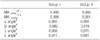
There was no significant difference in α, β, δ angle and mechanical axis between group I and II. There was a significant difference in the γ angle between group I and II (p<0.05). There was a significant difference in the α and β angle and mechanical axis between group IA and IB (p<0.05). There was a significant difference in the α angle and mechanical axis between group IIA and IIB (p<0.05). There was a positive correlation between the measured angle by the respective observers in all groups (p<0.05).
Conclusion
Patients with a preoperative varus deformity >20° tended to have more postoperative varus mechanical alignment than those with a preoperative varus deformity between 10° and 20° after TKA. More careful attention during the registration of the femoral mechanical axis should be paid in patients with a larger varus deformity in TKA using a computer-assisted navigation system. On the other hand, a reasonable mechanical valgus angle should be considered in femoral bone cutting for a varus deformity of the distal femur in conventional TKA. In addition, inadequate positioning of intramedullary rod should be recognized in conventional TKA.
Figures and Tables
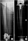 | Fig. 1(A) The femoral component angle (A) was measured between a line parallel to the femoral condyle and the mechanical axis of the femur on the anteroposterior view. (B) The tibial mechanical angle (B) was measured between a line parallel to the tibial condyle (a) and a line parallel to the ankle joint (b).
|
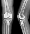 | Fig. 2Post-operative measurement of the femoral and tibial component. α, The inclination angle of the femoral component was formed with the anatomical axis of the femur. β, The inclination angle of the tibial component was formed with the mechanical axis of the tibial. γ, The angle of the femoral component was formed between the anatomical axis of the femur and perpendicular line to the prosthesis. δ, The angle of the tibial component was formed with the mechanical axis of the tibial.
|
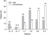
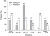
 | Fig. 4Comparison of the different distributions of the postoperative mechanical axis between Group IA and IB.
|
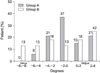
 | Fig. 5Comparison of the different distributions of the postoperative mechanical axis between Group IIA and IIB (ed note: I think the highlighted can be deleted).
|


References
1. Cates HE, Ritter MA, Keating EM, Faris PM. Intramedullary versus extramedullary femoral alignment systems in total knee replacement. Clin Orthop Relat Res. 1993. 286:32–39.

2. Engh GA, Petersen TL. Comparative experience with intramedullary and extramedullary alignment in total knee arthroplasty. J Arthroplasty. 1990. 5:1–8.
3. Garg A, Walker PS. Prediction of total knee motion using a three-dimensional computer-graphics model. J Biomech. 1990. 23:45–58.
4. Ishii Y, Ohmori G, Bechtold JE, Gustilo RB. Extramedullary versus intramedullary alignment guides in total knee arthroplasty. Clin Orthop Relat Res. 1995. 318:167–175.
5. Jiang CC, Insall JN. Effect of rotation on the axial alignment of the femur. Pitfalls in the use of femoral intramedullary guides in total knee arthroplasty. Clin Orthop Relat Res. 1989. 248:50–56.
6. Laskin RS. Instrumentation pitfalls: you just can't go on autopilot! J Arthroplasty. 2003. 18:18–22.
7. Lotke PA, Ecker ML. Influence of positioning of prosthesis in total knee replacement. J Bone Joint Surg Am. 1977. 59:77–79.
8. Mahaluxmivala J, Bankes MJ, Nicolai P, Aldam CH, Allen PW. The effect of surgeon experience on component positioning in 673 Press Fit Condylar posterior cruciate-sacrificing total knee arthroplasties. J Arthroplasty. 2001. 16:635–640.
9. Mielke RK, Clemens U, Jens JH, Kershally S. Navigation in knee endoprosthesis implantation--preliminary experiences and prospective comparative study with conventional implantation technique. Z Orthop Ihre Grenzgeb. 2001. 139:109–116.
10. Mihalko WM, Boyle J, Clark LD, Krackow KA. The variability of intramedullary alignment of the femoral component during total knee arthroplasty. J Arthroplasty. 2005. 20:25–28.
11. Moreland JR. Mechanisms of failure in total knee arthroplasty. Clin Orthop Relat Res. 1988. 226:49–64.
12. Oswald MH, Jakob RP, Schneider E, Hoogewoud HM. Radiological analysis of normal axial alignment of femur and tibia in view of total knee arthroplasty. J Arthroplasty. 1993. 8:419–426.
13. Reed SC, Gollish J. The accuracy of femoral intramedullary guides in total knee arthroplasty. J Arthroplasty. 1997. 12:677–682.
14. Ritter MA, Faris PM, Keating EM, Meding JB. Postoperative alignment of total knee replacement. Its effect on survival. Clin Orthop Relat Res. 1994. 299:153–156.
15. Sambatakakis A, Wilton TJ, Newton G. Radiographic sign of persistent soft-tissue imbalance after knee replacement. J Bone Joint Surg Br. 1991. 73:751–756.
16. Seon JK, Song EK. The accuracy of lower extremity alignment in a total knee arthroplasty using computer-assisted navigation system. J Korean Orthop Assoc. 2004. 39:566–571.
17. Sparmann M, Wolke B, Czupalla H, Banzer D, Zink A. Positioning of total knee arthroplasty with and without navigation support. A prospective, randomised study. J Bone Joint Surg Br. 2003. 85:830–835.
18. Stulberg SD, Loan P, Sarin V. Computer-assisted navigation in total knee replacement: results of an initial experience in thirty-five patients. J Bone Joint Surg Am. 2002. 84:Suppl 2. S90–S98.
19. Tang WM, Chiu KY, Kwan MF, Ng TP, Yau WP. Sagittal bowing of the distal femur in Chinese patients who require total knee arthroplasty. J Orthop Res. 2005. 23:41–45.
 XML Download