

 PDF
PDF ePub
ePub Citation
 Print
Print


INTRODUCTION
Hypoglycemia is caused by imbalance between glucose supply and glucose utilization. It is apt to be caused in diabetic patients as a side effect of excessive insulin and sulfonylurea uses. When hypoglycemic injury affects brain tissue and induces neurologic abnormalities, it is called hypoglycemic encephalopathy.
There is a report of DWI image changes from subcortical white matter lesions to cortical lesions in hypoglycemic encephalopathy, but it took several days for the changes (1). We present a case of hypoglycemic encephalopathy, showing rapid changes and uncommon findings on sequential MRI examinations.
CASE REPORT
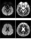
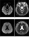
A 58-year-old male patient with a history of diabetes presented with semicoma status. He was alert as usual, just 1 hour before being found in unresponsive status. On arrival at emergency department, vital signs were stable. His initial blood glucose level was 44 mg/dL and no other abnormality was found in his blood test. 10% D/W and 50% dextrose were continuously infused and blood glucose level went up as high as 292 mg/dL but his mental status was unchanged. After the rapid correction of blood glucose level for an hour, initial brain MRI was done immediately and it showed bilateral subcortical white matter lesions, which was hyperintense on diffuse DWI, hypointense on apparent diffusion coefficient (ADC) (Fig. 1). Deep and cortical gray matters were spared. He had no history of stroke and traumatic brain injury and no other abnormal findings on arterial blood gas analysis (ABGA), vital signs, and electrolyte. There was no other possible cause for his sudden mental change with abnormal findings on MRI. Approximately 5 hours 3 mins after the initial MRI scan, follow-up MRI scan was performed at the emergency department. Follow-up MRI showed resolved subcortical white matter lesions, with newly-appeared DWI high signal intensity lesions in bilateral fronto-temporo-parietal cortex (Fig. 2). He had admitted to the hospital for 3 more days and transferred to another hospital on his family's will. During the hospitalization, the blood glucose level was maintained within normal limits and no other causative brain damage was noted but his mentality remained unchanged.
DISCUSSION
Glucose is known as mainstay of brain energy source. Therefore profound glucose deficiency may lead to neuronal necrosis, which is very identical to neuronal ischemic damage by oxygen deficiency, and it has been demonstrated in pathologic studies (2). Glucose deprivation may lead to brain energy failure, membrane ionic pump failure and tissue alkalosis, causing a shift of water into the intracellular space and a drastic reduction of extracellular space volume and leading to selective neuronal necrosis, especially in the cerebral cortex, basal ganglia, and hippocampus (3).
Various patterns of hypoglycemic encephalopathy are shown on DWI (4). But, the mechanism of ADC reduction in hypoglycemia is still unclear. Cortical laminar necrosis may be the most likely mechanism for the hyperintensity revealed by DWI during subacute period (5). In an animal study, Hasegawa et al documented global cortical decline in ADC rapidly spreading to the entire brain, showing neuronal necrosis in the hippocampus and the neocortex in animals with prolonged hypoglycemia. But the reductions in ADC were transient and were reversed by glucose infusion (6). Reversibility of the ADC abnormalities can be explained by the mechanism of excitotoxic edema. Increased extracellular glutamate causes calcium and sodium entering into cells, inducing apoptosis, however, glutamate also induces edema of glial cells and myelin sheaths, called excitotoxic edema, which might protect axons from irreversible damage (7). Moreover, the receptors related to excitotoxic mechanisms are widely distributed in both gray and white matter, which may explain the widely distribution of early DWI abnormalities (8). Diffuse and extensive injury observed on DWI predicts a poor neurologic outcome in hypoglycemic encephalopathy (4).
In patients with hypoglycemic encephalopathy, brain parenchymal lesions were typically shown to be distributed bilaterally in the basal ganglia, hippocampus, cerebral cortex, and/or substantia nigra but not in the thalamus (9). However, this case shows a DWI of hypoglycemic encephalopathy patient presented with subcortical white matter lesions followed by bilateral fronto-temporo-parietal cortical lesions. Recently, there were several reports showing hypoglycemic encephalopathy patients with an untypical finding of diffuse white matter changes (4, 10). Another case report showed that hypoglycemic encephalopathy patient presented with subcortical white matter lesions and 14 days later demonstrating new hyperintense lesions involving bilateral cerebral cortex while previous subcortical white matter lesions disappeared (1). But to the best of our knowledge, this is the first case report in which white matter lesions rapidly resolve and cortical lesions follow just in about 5 hours.
The explanations for these rapid changes from white matter lesions to gray matter lesions remain unknown. It has been proposed that brain tissue have different vulnerability to hypoglycemia, therefore, gray matter showed delayed presentation of DWI abnormalities (1). We proposed that variable factors, such as severity of hypoglycemic damage or individual difference in vulnerability to hypoglycemia, might determine the time interval between the changes of white matter and gray matter lesions. But these hypotheses remain yet to be further explained.
Our report has a limitation that we are not sure of what sort of injury had basically induced brain damage to our patient. But considering he had been imprisoned for several days and no other possible causative agents were found in prison, plus, he only demonstrated low blood glucose level in physical exam and laboratory tests, hypoglycemia had most likely induced both bilateral cortex and white matter injuries.
In conclusion, we described the neurochemical pathophysiology of hypoglycemic encephalopathy, the mechanism of DWI changes in hypoglycemia and some hypotheses for the rapid changes from white matter lesions to gray matter lesions. It is important to know that MR imaging changes in hypoglycemic encephalopathy can be made as quick as just a few-hour-long, quicker than we have known through the previous report. Performing early follow-up DWI scan might be helpful in quickly determining the extent of brain damage and quickly accessing the clinical severity in some patients with hypoglycemic encephalopathy.
 XML Download