

 PDF
PDF ePub
ePub Citation
 Print
Print


INTRODUCTION
Syphilis is a sexually transmitted disease caused by a spirochete, Treponema pallidum (T. pallidum). Involvement of the central nervous system (CNS) occurs in 5-30% of the patients and the CNS may be affected at any stage of the disease (1). It is difficult to differentiate neurosyphilis from other cerebral infectious and inflammatory diseases, because of its various times of onset and diverse form of presentation. Major forms of neurosyphilis include asymptomatic, acute syphilitic meningitis, meningovascular syphilitis, parenchymal and gummatous neurosyphilis (2). A reactive cerebrospinal fluid (CSF) venereal disease research laboratories (VDRL) at any stage of syphilis is required for confirmation of neurosyphilis; however, the test is negative in approximately half of the patients and the sensitivity of CSF VDRL is relatively low (3). Therefore, magnetic resonance imaging (MRI) may help to make an earlier diagnosis of neurosyphilis in a patient with syphilis and neurologic symptoms.
Neuroradiological findings regarding neurosyphilis, particularly MR imaging, are varied and CNS abnormalities are nonspecific edematous or enhancing brain parenchymal lesions, meningitis, neuritis, and cerebral infarction, and are rarely spinal cord lesions (1, 2, 4, 5, 6). Herein, we report a case of both meningovascular and spinal meningomyelitic types of neurosyphilis in a human immunodeficient virus (HIV)-negative patient, which involved multiple cranial nerves, large to medium size cerebral arteries and the spinal cord.
CASE REPORT
A 53-year-old man was admitted to neurology department complaining of headache, dizziness and blurred vision. Right side hearing disturbance and right facial palsy had developed three months before admission. The patient had been diagnosed with Ramsay-Hunt Syndrome at that time, and was treated via an anti-viral agent and steroids. Nine months before admission, he had visited the urology clinic for a painless ulcerative penile mass, a penile gumma. At that time, his serum VDRL titer was 1:128, and fluorescent treponemal antibody absorbed (FTA-ABS) test IgG was reactive. Antigen and antibody for HIV were negative. The penile lesion had spontaneously regressed and he was absent for follow ups over several months.
At admission, bitemporal hemianopsia and right side sensorineural hearing loss were demonstrated on a visual field examination and an audiogram, respectively. He was alert without any evidence of cognitive or memory disturbance, and other neurologic tests, including his sensory or motor functions were normal. In laboratory tests, his serum VDRL titer was 1:20, and FTA-ABS test IgG was reactive. A CSF examination revealed lymphocyte-predominant pleocytosis (6 nucleated cells/mm2 [91% lymphocytes]). His CSF protein was elevated (119 mg/gl, normal range; 12-60 mg/gl). No microorganisms were found in the CSF.
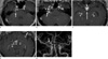
Brain MRI revealed diffuse leptomeningeal enhancement on postcontrast T1-weighted images, representing meningitis (Fig. 1). There were multiple enhancing lesions along the right internal auditory canal, right hypothalamus, pituitary stalk, right Meckel's cave and Gasserian ganglion, intracranial segments of both optic nerves and chiasm. Enhancements of right cochlea and vestibule were also present. In addition, multifocal thick, irregular and nodular enhancing lesions were observed in the basal cistern and left Sylvian fissure. Time-of-flight and contrast enhanced magnetic resonance angiography (MRA) revealed a segmental stenosis at the M1 portion of the right middle cerebral artery. Because of the patient's visual symptom, spine MRI was performed to rule-out the possibility of demyelinating diseases, such as neuromyelitis optica. A segmental T2 high signal intensity was observed at the central portion of the spinal cord at the T7 level. Postcontrast T1-weighted images revealed incomplete patchy enhancement in the region of the cord with T2 high signal intensity. A nodular meningeal enhancement along with enhancing clumped nerve roots was observed at the L1 level. Diffuse meningeal enhancement along the surface of the lumbar and lower thoracic cord and the cauda equine was also noted. However, there was no corresponding neurologic symptom. (Fig. 2).
According to the MRI findings, meningovascular and the spinal form of neurosyphilis was strongly considered in this patient. Further laboratory tests were performed, and a reactive CSF VDRL test (titer, 1:8) was obtained. The patient was finally diagnosed with neurosyphilis, and Penicillin G benzathine was intramuscularly administered at a rate of 2.4 million units over 14 times. A follow-up MRI after 12 days demonstrated improved enhancing lesions along the 2nd, 5th, 7th, and 8th cranial nerves and we also noted an improved meningeal enhancement. The degree of enhancement of right membraneous labyrinth was also decreased. However, an acute infarction was found in the left centrum semiovale on diffusion-weighted imaging, and MRA revealed multifocal segmental stenosis in the left proximal middle and posterior cerebral arteries. Two weeks after admission, his visual symptoms improved. One months after completion of the antibiotic therapy, his optic, trigeminal, facial and vestibulocochlear nerve functions improved and his serum VDRL titer decreased (1:8). A follow-up spinal MRI was not performed because he had no related neurologic symptoms.
DISCUSSION
Syphilis is a sexually transmitted disease caused by the spirochete bacterium Treponema pallidum (T. pallidum). The clinical course of syphilis is divided into the following three stages; primary, secondary, and tertiary syphilis. The primary stage is characterized by a round painless sore, a chancre, at the inoculation site. Hematologic dissemination and the secondary syphilis occur within 2 to 4 weeks, if untreated. This clinical stage typically manifests as a rash on the palms of the hands and soles of the feet, and the CNS can be involved in the secondary stage observed as aseptic meningitis. Tertiary syphilis usually appears 5 years or more after the primary infection. Tertious syphilis includes cardiovascular syphilis, neurosyphilis, and late benign syphilis as gummatous syphilis (4). The prevalence of syphilis was significantly reduced after the development of penicillin. However, in the last few decades, cases of syphilis have increased as the incidence of HIV infections has increased. As a result, the incidence of neurosyphilis may have also increased. Neurosyphilis is a slow progressive infection of the brain and spinal cord. Neurosyphilis is seen primarily in the tertiary and occasionally in the secondary stages of the disease (1, 2, 4). Clinically symptomatic infections are categorized into acute syphilitic meningitis, meningovascular syphilis, parenchymatous and gummatous syphilis. Spinal involvement is an extremely rare presentation (5, 6, 7). The MRI features of neurosyphilis vary as do the clinical presentations. In this case report, we described the brain and spinal MRI findings of neurosyphilis in an HIV-negative patient with multiple cranial nerve palsy and a cerebral infarction. The symptoms and imaging findings in our patient were most consistent with those seen in meningovascular syphilis involving the basilar meninges and cranial nerves and syphilitic meningomyelitis (2, 5, 6, 7, 8, 9, 10). Brain and spine MRI revealed extensive cranial nerve involvement (the 2nd, 5th, 7th, 8th cranial nerves), proximal cerebral arteritis, and meningomyelitis of spine. To the best of our knowledge, a case presenting with both meningovascular and spinal form is extremely rare.
A wide range of brain imaging findings may be seen in neurosyphilis, and many of the clinical neurosyphilis have no demonstrable imaging abnormalities (8). The CNS is involved early in the course of the disease, and it is assumed that T. pallidum is cleared from the CNS in most patients (11). Failure of the immune system to clear the organism is thought to result in neurologic manifestations. Atrophy, cerebral infarction, white matter lesions and enhancing nodules referred to as gummas are included in the parenchymal form of neurosyphilis. The parenchymatous type is a well known CNS manifestation of tertiary syphilis and results in general paresis secondary to widespread parenchymal damage and tabes dorsalis secondary to demyelination of the posterior columns, dorsal roots, and dorsal root ganglia (4, 8). Meningovascular syphilis is less frequent and may produce cerebral infarction, meningoneuritis, meningolabyrinthitis or labyrinthitis (8). The symptoms present within months to as late as a decade after primary infection (8). Enhancement of the oculomotor, trigeminal, facial, vestibulocochlear, glossopharyngeal, vagal and spinal accessory nerves has been reported, and neural enhancement is likely secondary to a breakdown of the blood-brain barrier secondary to the inflammation (6, 8, 9, 10). Enhancement of cranial nerves and meninges may be seen in a broad range of pathologic entities, including neoplastic, granulomatous, infectious, and posttraumatic diseases. The extensive involvement of the cranial nerves, meninges and membraneous labyrinths is unlikely seen in primary or metastatic tumors, and other infectious diseases such as herpes zoster or tuberculosis. In this report, various manifestations of meningovascular neurosyphilis presenting as meningoneuritis involving multiple cranial nerves (the 2nd, 5th, 7th, 8th cranial nerves), meningolabyrinthitis, cerebral arteritis, were discovered in a single patient.
Spine involvement is an extremely rare manifestation of neurosyphilis, and a few reports have described the MRI features of noncompressive myelopathy, and subacute meningomyelitis (5, 6, 7). Myelitis, in the form of either segmental or intrinsic spinal cord signal changes in the thoracic region on T2-weighted images, along with a patchy enhancement after gadolinium enhancement has been described in the literature (5, 6). Syphilitic meningomyelitis had been described as an enhancement along the cauda equine and ventral surface of the spinal cord with extensive cord swelling and diffuse high signal intensity on T2-weighted imaging (7). The spinal lesions seen in our case might be considered in the diagnosing syphilitic meningomyelitis, yet, there was no representative neurologic symptom in the patient. In the literature, the T2 hyperintense spinal cord lesions and contrast enhancement were reversible after the administration of penicillin G, although the spine MRI abnormalities were not followed in our patient, because he had no related neurologic symptom (5, 6, 7).
The positive result of the CSF VDRL test and the rapid response of cranial nerve dysfunction to penicillin G therapy confirmed the diagnosis of neurosyphilis in this patient. Because a CSF VDRL test is not routinely performed in patients who have syphilis and because the sensitivity of this test is relatively low (3, 4), a clinical inspection of neurosyphilis should be given in patients with multiple cranial nerve dysfunction, based on serologic tests, CSF analysis and MRI findings.
In conclusion, this patient showed diverse brain and spine MRI findings of meningovascular neurosyphilis and syphilitic meningomyelitis. He was successfully treated with antibiotics after an appropriate diagnosis. Although the incidence of neurosyphilis is rare, the MRI findings of the present case suggest that neurosyphilis should be considered in the differential diagnosis of a challenging case which had combined meningoneuritis, cerebral infarction and meningomyelitis.

 XML Download