

 PDF
PDF ePub
ePub Citation
 Print
Print


INTRODUCTION
Wandering spleen is a rare clinical condition characterized by ectopic position of the spleen due to abnormal peritoneal attachments including the lienorenal and gastrosplenic ligaments (1). Wandering spleen and intestinal malrotation share a common cause: the absence of an intraperitoneal visceral ligament (2). Wandering spleen presents a wide range of clinical manifestations, from incidental finding to life-threatening conditions. The laxity of such suspensory ligaments of spleen predisposes torsion of the splenic vascular pedicle, leading to splenic infarction (3). These fatal complications, however, are not characterized by specific clinical symptoms; hence, image evaluation is crucial for detecting such complications. Radiologic imaging features of wandering spleens have been reported in previous studies, but to our knowledge, only a few cases of infarcted wandering spleen were associated with intestinal malrotations (2, 4). Herein we describe MR images of an unusual case of infarction of wandering spleen in association with intestinal non-rotation in a pediatric patient.
CASE REPORT
A 7-year-old girl was referred to our hospital after an abdomen CT that revealed a mass in the lower abdominal cavity. She had a 3-day history of abdominal pain, diarrhea, and vomiting. She was afebrile and her vital signs were stable on admission. Physical examinations revealed tenderness in the mid-lower abdomen without rebound tenderness or palpable mass. Laboratory evaluations revealed a high white blood cell count of 14.7 K/µL with 76% of neutrophils, hemoglobin of 11.7 g/dL, hematocrit of 35.7%, platelets of 354 K/µL and elevated serum C-reactive protein of 11.9 mg/dL.
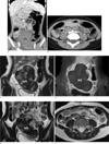
In plain radiography, large mass-like opacity was observed in the right-lower abdomen, and gas of ascending and transverse colon was pushed to the left side. Contrast-enhanced abdominal CT scan revealed a 6.5×6.3×9.6 cm (transverse×AP×craniocaudal dimension) non-enhancing soft tissue mass in the lower abdominal and pelvic cavity (Fig. 1a). Whirling splenic vessel with weak enhancement and perivascular edema was demonstrated (Fig. 1b). We suspected that the soft tissue mass was a wandering spleen with infarction due to vascular compromise. The large bowel was predominantly on the left side of the abdomen and the small bowel was on the right side of the abdomen, indicating non-rotation of gastrointestinal tract. T2-weighed images (slice thickness = 5 mm, FOV=180×180, Matrix = 352×192, no gap, TR/TE=4883/104) revealed heterogeneous signal intensity in wandering spleen and accessory spleens (Fig. 1c, e). Contrast-enhanced (Gadodiamide, 0.1 mmol/kg, IV, Omniscan, GE healthcare) fat suppressed T1-weighted images (slice thickness=5 mm, FOV=180×180, Matrix = 352×192, no gap, TR/TE=666/11) showed non-enhancing infarcted wandering spleen with enhancing foci near the splenic hilum (Fig. 1d). Twisted or whirling splenic vessel was more evidently seen as signal void, in contrast to high signal intensity of perivascular edema, omental fat (Fig. 1e, f).
Thus, laparoscopic excision of the wandering spleen was performed. On laparoscopy, the wandering spleen was 12.0 cm in diameter and torsion of the splenic vessels was demonstrated. In the upper-left quadrant, three non-infracted accessory spleens were observed. Intestinal non-rotation was demonstrated as the right-sided small bowel loops and the left-sided colon. Grossly, the capsule of the spleen was purplish red and intact. Histological examination confirmed total infraction of the wandering spleen. The postoperative course was uneventful, and the patient was discharged on the 5th day after the operation.
DISCUSSION
Wandering spleen is, by definition, a mobile spleen that is attached only by an elongated vascular pedicle, causing the spleen to migrate to any part of the abdomen or pelvis (2). If the pedicle is twisted in the course of movement of the spleen, blood supply may be interrupted or blocked, resulting in severe damage to the blood vessels (5). Acute splenic torsion compromises venous outflow, which causes congestion and impairment of arterial inflow. Pain is originated from the splenic capsular stretching with rapid splenic enlargement and localized peritonitis (6).
During fetal development, failure to fuse with the posterior peritoneum leads to a laxity or absence of formation of splenic supporting ligaments including pancreaticocolic, splenocolic, gastrosplenic, pancreaticosplenic, phrenicocolic, splenorenal, and phrenicoscplenic ligaments (2). Although wandering spleen and intestinal malrotation are uncommon and can occur independently, these entities share a common congenital etiology: anomalous intraperitoneal visceral attachment that originates from the dorsal mesentery (4).
There are multiple causes of this ligament laxity including congenital and acquired etiologies. The former indicates failure of development of the normal splenic suspensory ligaments including the lienorenal and gastrosplenic ligaments (1, 2). The acquired form can occur under conditions that weaken the ligaments including pregnancy with elevated estrogen levels and abdominal wall laxity after trauma (2, 7). As the spleen normally arises from fusion of splenucluli from the dorsal mesogastrium, it is unlikely that gut malrotation and wandering spleen affect each other.
By ultrasound, acute torsion of wandering spleen is seen as an ectopic comma-shaped spleen, and no orthotopic splenic tissue is found in the upper-left quadrant (8). Lack of demonstrable flow within the splenic parenchyma, both in the color flow survey and directed search with duplex Doppler; and possible alterations in main splenic arterial flow, specifically, elevation of the resistive index or other measures of vascular impedance (8). Contrast-enhanced CT preferred to diagnose a wandering spleen when torsion is suspected clinically or is suggested by other imaging studies. Minimal, patch, or no enhancement of spleen was a finding that strongly indicates compromised splenic perfusion (3). Mild enhancement may be attributed to collateral circulation through the short gastric and left gastric veins and/or the short gastric and pancreatic arteries. Perhaps the most specific sign of torsion is a whirly appearance of splenic vessels and surrounding fat, usually noted at the splenic hilum (3, 9).
MRI can easily reveal lack of splenic tissues in the left upper quadrant the ectopic location of spleen in pelvic cavity on T1- and T2-weighted sequences. The viability of the splenic parenchyma can be assessed by T1-weighted images with contrast medium administration (10). In MRI, compared to in CT, tortured vascular pedicles are seen as signal void, contrary to a high signal intensity of perivascular fat, evidently. This advantage could help distinguish twisted vascular structure from omental fat (11).
Detorsion, splenopexy and splenectomy are the treatment options for children with wandering spleens. Because of the risk of post-splenectomy sepsis, surgeons prefer to maintain the organ with splenopexy, except for in patients with massive splenic infarction or splenic vessel thrombosis (12). A variety of methods of performing splenopexy were described: direct suture of capsule or hilum, peritoneal pocket, absorbable mesh, and synthetic mesh (13). Prophylatic gastropexy in patients with wandering spleen is usually the treatment of choice when gastric volvulus is present (2, 4).
In summary, it is important to recognize a wandering spleen and intestinal malrotation because of configuration of vascular pedicle or peritoneal laxity, which makes it prone to splenic torsion and/or intestinal volvulus. These entities are not characterized by specific symptoms, so imaging diagnosis of these complications plays an important role for immediate surgical management. CT is a popular way to diagnose wandering spleens or associated abnormalities, and has a short scan time, which is advantageous for patients who cannot hold position for long. But the radiation from CT is hazardous for pediatric patients. Thus, MRI can be a safe alternative for diagnosis of wandering spleen for young patients. Also, compared with CT scan, MR image contrast between the signal-void vascular pedicles and the hyperintense perivascular fat tissue is an advantage for diagnosis of vascular torsion.
 XML Download