

 PDF
PDF ePub
ePub Citation
 Print
Print


INTRODUCTION
Primary non-Hodgkin lymphoma (NHL) of the breast is a rare clinical entity, representing about 0.4-0.5% of all breast malignancies and 1.7-2.2% of all extranodal NHL (1, 2, 3). We describe two cases of primary breast lymphomas in a middle aged woman and discuss characteristics of classical findings of classical magnetic resonance (MR) imaging, MR spectroscopy and diffusion weighted (DW) imaging as well as mammography and ultrasonography.
CASE REPORTS
Case 1
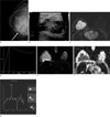
The 48-year-old woman visited our hospital with a palpable mass on her right breast which has been increasing in size for the past two months. Physical examination revealed a hard and non-tender mass in the right breast. There were no signs of nipple retraction or skin thickening. Mammography demonstrated a large mass on the right breast with oval shape, circumscribed margin, and high density (Fig. 1a). On ultrasonography, the mass showed an oval shape, parallel orientation, circumscribed margin, heterogeneous echogenicity, and no posterior features (Fig. 1b). The fat-suppressed T2-weighted MR images showed an irregular circumscribed mass that was slightly hyperintense relative to parenchyma. On the contrast-enhanced MR imaging, a mass measuring 8.1 cm was observed in the right breast (Fig. 1c). It was an irregular shape and circumscribed margin with homogeneous enhancement. On dynamic imaging, the lesion displays fast contrast uptake in the early phase and rapid washout in the delayed phase consistent with type III kinetic curve (Fig. 1d). DW imaging (b=1000) showed reduced diffusion with corresponding mean apparent diffusion coefficient (ADC) value of 0.75 × 10⁻³ mm2/s (Fig. 1e and f). MR spectroscopy showed a prominent Cho peak at 3.22 ppm, which was consistent with malignancy (Fig. 1g). The MR spectroscopy imaging was performed on a 3 tesla machine (Verio, Simens, Erlangen, Germany). Choline signal alterations associated with tumor were quantified using the 'normalized choline' signal approach. Using normalization, the choline signal can be calibrated to a relative and standardized value automatically. To perform this choline signal quantification, the choline signal in the tissue of interest is measured as is the choline signal generated by external reference probe. The specific imaging parameters were as follows: TR/TE 1500/100, number of excitations 128, sequence acquisition time 3 min 18 sec. Breast biopsy performed for histopathological identification of the mass was diagnostic for diffuse large B-cell lymphoma (CD 20+). Laboratory tests, PET/CT scan, CT of chest and abdomen, and bone marrow biopsy showed no involvement of other sites.
Case 2
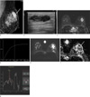
The 40-year-old woman presented with 3-week history of a painless left breast lump. On physical examination, a hard mass was palpated on the left breast. There were no other clinical symptoms such as nipple retraction, nipple discharge, or skin thickening. Mammography demonstrates a 4 cm mass on the left breast with oval shape, circumscribed margin, and equal density (Fig. 2a). Breast ultrasound showed an oval, parallel oriented mass with circumscribed margin at the 12 o'clock position of the left breast (Fig. 2b). It showed heterogeneous echogenicity with posterior enhancement. On the fat-suppressed T2-weighted MR images, the mass showed an irregular shape, circumscribed margin, and slightly hyperintense relative to parenchyma. The mass showed strong homogeneous internal enhancement after contrast material injection on MR imaging (Fig. 2c). On dynamic imaging, the lesion showed fast contrast uptake in the early phase and persistent enhancement in the delayed phase consistent with type I kinetic curve (Fig. 2d). DW imaging showed reduced diffusion with corresponding mean ADC value of 0.82 × 10⁻³ mm2/s (Fig. 2e and f). High-amplitude Cho peak at 3.22 ppm is detected by single voxel MR spectroscopy (Fig. 2g). Ultrasound guided core needle biopsy performed on her breast mass and the histopathology showed diffuse large B-cell lymphoma after immunohistochemistry (CD 20+). Laboratory tests, PET/CT scan, CT of chest and abdomen, and bone marrow biopsy showed no involvement of other sites. The patient was treated with chemotherapy.
DISCUSSION
Primary breast lymphomas is a rare disease accounting for less than 0.5% of all breast malignancies and this rarity may be related to paucity of the lymphoid tissue in the breast (2, 3) The diagnosis of primary breast lymphoma is limited to the breast which is the first or major manifestation site of lymphoma, and there is no evidence of disseminated lymphoma other than simultaneous ipsilateral axillary node involvement, and no prior diagnosis of lymphoma (2). Most breast lymphomas are of B-cell origin with a predominance of diffuse large cell lymphoma and, rarely, of T-cell or histiocytic lymphoma (2). The manifesting symptoms are founded mostly in palpable masses. And, they are sometimes painful and generally located located in the upper outer quadrants (1). B symptoms are uncommon. Similar to previous studies, our patients presented to the hospital with complaints of a palpable breast mass.
The imaging finding of primary breast lymphoma is nonspecific (4, 5, 6). Appearances on mammography are variable but mostly present as circumscribed round to oval dense and noncalcified mass (4). Similar to previous reports, our cases revealed an oval, circumscribed, and high density mass on mammography. Architectural distortion or diffuse increased parenchyma density with or that without skin thickening was also reported (4, 6). There is no specific mammographic finding that would differentiate primary breast lymphoma from other invasive carcinomas of the breast. On ultrasonography, the most common feature of primary breast lymphoma was a solitary hypoechoic, homogeneous, or heterogeneous circumscribed mass (4). In addition, these lesions are often significantly hypoechoic that they can be mistaken for simple cyst (4). In our cases, the mass shows an oval, circumscribed, and heterogeneous echogenicity in ultrasonography. This finding agrees with data presented in previous reports. However, a broad range of sonographic appearance, ranging from circumscribed to ill-defined margins, hypoechogenicity to hyperechogenicity, focal or diffuse involvement has been reported previously (4, 5).
There are a few reports defining MR imaging findings of primary breast lymphoma (7, 8, 9). They are reported as a non-spiculated, isointense mass on T1-weighted images, and a hyperintense mass on T2-weighted images. On dynamic imaging, the mass shows homogeneous rapid and strong enhancement which indicates malignant process. Homogeneous enhancement on dynamic images differentiates malignant lymphoma from invasive breast carcinoma of no special type which shows heterogeneous internal enhancement or rim enhancement in most cases. The homogeneous enhancement may reflect the uniform proliferation of the tumor cells. However, because the MR findings of breast lymphoma are not pathognomonic, biopsy is essential for definite diagnosis.
All of the described kinetic curve can occur in breast lymphoma: type I with persistent enhancement; type II with plateau pattern; type III with a washout enhancement pattern. According to the previous reports, type II kinetic curve was present in most cases. One case shows type I kinetic curve and another case shows type II kinetic curve in this study.
DW imaging is a specific modality that visualizes the microstructural characteristics such as tissue cellularity, which has been shown to be an important index of tumor grade, and local tissue architecture, which is a sensitive early indicator of abnormality (10). Restricted water diffusion in tumors with increased cell density and reduced extracullular space usually leads to higher signal intensity and decreased ADC (10). The ADCs of malignant breast lesions are usually lower than those of benign lesions, indicating restricted water diffusion and increased cellularity (11). Matsubayashi et al. (12) reported that the primary breast lymphoma shows homogeneous very high signal intensity on DW imaging, which may reflect the high cellular density of the tumors. This finding was similar to those observed in our cases.
MR spectroscopy is a noninvasive technique for the assessment of biochemical tissue properties that can be readily incorporated into standard diagnostic MR imaging protocols. The method reflects the chemical composition of cells and the changes that occur with tumor development and progression (13, 14). Increased levels of total Cho have been detected in malignancies and are ascribed to an increased cellular membrane turnover (13, 14). The results of several MR spectroscopy studies have shown elevated levels of Cho in the majority of breast carcinomas (15). To the best of our knowledge, the MR spectroscopy of primary breast lymphoma has been described in only one report in the English medical literature (16). According to the report, MR spectroscopy findings of primary breast lymphoma show elevated Cho resonance peak. These findings are consistent with the malignancy, which is similar to our cases.
In summary, primary lymphoma of the breast is often a challenge to diagnose because of disease rarity and nonspecific imaging findings. Primary lymphoma of the breast in advanced MR imaging appears as reduced diffusion on DW imaging and elevated Cho peak on MR spectroscopy in our cases. As in our cases, a combination of various modalities and techniques is helpful to diagnose this entity from primary breast carcinoma or even benign breast tumor. Although breast MR does not provide specific information for the diagnosis of lymphoma, advanced sequences including DW imaging and MR spectroscopy can provide more clues for differentiation from benign or malignant lesions.
 XML Download