

 PDF
PDF ePub
ePub Citation
 Print
Print


INTRODUCTION
Endophthalmitis refers to an intraocular inflammatory process that may result from exogenous (primary, postoperative, post-traumatic, infection) or endogenous causes (1). Endogenous endophthalmitis is rare, accounting for 2 to 8% of all case of endophthalmitis and results from hematogenous bacterial spread (1).
Staphyloccocus aureus is the most common pathogenic cause of bacterial endogenous ophthalmitis in the West (1), while Klebsiella pneumoniae is the most common pathogen in the East (2). In the Western literature, the most common non-ocular source of infections are endocarditis, urinary tract infection, meningitis, and wound infection. In contrast, most cases of endogenous endophthalmitis in the East arise from liver abscesses (1-3).
We report a case of simultaneously developed endogenous endophthalmitis and brain abscesses, especially focused on magnetic resonance imaging (MRI) findings.
CASE REPORT
A 55-year-old man with poorly controlled diabetes mellitus was admitted to a local hospital for the management of upper respiratory infection symptoms and nonsense talking. His illness was diagnosed as bacterial meningitis by cell count analysis of cerebrospinal fluid (CSF). The patient's symptoms improved during antibiotic treatment. However, three days before transfer to our hospital, the patient developed blurred vision in his right eye, which rapidly progressed to blindness. He was transferred to our hospital, where an MRI scan was performed.
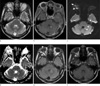
A T2-weighted image (GE Signa EXCITE 1.5T MRI; TR 4,000 ms; TE 106 ms; FOV 210 × 210 mm; slice thickness 5 mm) showed multiple hyperintensities in both cerebral hemispheres, all of which had rim enhancement visible on gadolinium-enhanced T1-weighted images (TR 700 ms; TE 12 ms, Fig. 1a, b). Almost of above-mentioned hyperintensities were observed on diffusion-weighted image (DWI, b value 1000s/mm2) with corresponding hypointensity on apparent diffusion coefficient (ADC) mapping (Fig. 1c, d). Hyperintensity was observed in the right globe in DWI and fluid attenuated inversion recovery (FLAIR, TR 8000 ms; TE 140 ms; TI 2500 ms; flip angle 90 degree) images, with corresponding hypointensity on ADC mapping (Fig. 1c, d, e). A T1-weighted image (TR 600 ms; TE 11 ms) showed subtle heterogeneous signal intensity in the right globe (Fig. 1f).
Laboratory tests were negative for CSF and urine cultures; however, a blood culture showed growth of Klebsiella pneumoniae. Based on these results, a provisional diagnosis of endogenous endophthalmitis resulting from a Klebsiella pneumonia-induced brain abscess was made. Seven days after aggressive intravenous antibiotics treatment, the patient's condition improved, although the blindness in the right eye persisted. We recommended an abdominal CT for evaluation of a liver abscess, but he refused. The patient was discharged three days after admission.
DISCUSSION
MRI is a useful imaging modality to diagnose endophthalmitis, with DWI, ADC and FLAIR being particularly effective. Because of globe hyperintensity on DWI and corresponding hypointensity on ADC mapping are suggesting intra-ocular or subretinal abscess formation (4), they are useful factors for the diagnosis of endogenous endophthalmitis, and hyperintensity on FLAIR imaging also suggests endogenous endophthalmitis. Although the best imaging tool for diagnosis of brain abscess (5), gadolinium-enhanced T1-weighted imaging is useless in the diagnosis of endophthalmitis because of the vitreous humor is avascular and an intravitreous abscess may not produce an enhancing wall before the occurrence of inflammatory neovascularization (4, 6). In the current case, as mentioned above, DWI, ADC and FLAIR sequences were useful for diagnosing endophthalmitis, and DWI and ADC were also valuable to reach the diagnosis of brain abscess. To our knowledge, imaging findings of bacterial endophthalmitis with brain abscess have been reported several times in English (7-9). However, this is the first report of imaging findings of simultaneously developed bacterial endophthalmitis with brain abscess on DWI and ADC. Therefore, this report will be a valuable case to illustrate the usefulness of DWI and ADC sequences for the diagnosis of simultaneously developed bacterial endophthalmitis with brain abscess.
 XML Download