

 PDF
PDF ePub
ePub Citation
 Print
Print


INTRODUCTION
According to the most recent WHO classification (2002 edition), soft tissue malignant fibrous histiocytoma (MFH) is considered to be an undifferentiated
pleomorphic sarcoma. MFH is the most common somatic sarcoma involving deep soft tissues (1). The most frequent primary sites of
MFH are the lower limbs (49%), upper limbs (19%), and abdominal cavity or retroperitoneum (16%) of adults (2). MFH of the breast is extremely rare, only 65 cases have previously been reported so far (3). And most of the
case reports were about histopathological diagnosis of primary breast MFH or therapeutic strategy and prognostic factors (3). In
recent study, there was an attempt to describe the imaging feature of primary breast sarcoma (4), but including only one breast
MFH among 24 breast sarcoma cases. And individual imaging findings of the specific types of breast sarcoma were not separately described in this study (4). Furthermore most of the reports of breast MFH are focused on primary breast MFH, so little is known about metastatic breast MFH. In
this case report, we describe imaging findings of metastatic breast MFH from left upper extremity, including breast MRI and other breast imaging modalities such as
mammography and ultrasound.
CASE REPORT
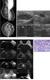
A 79-year-old woman was diagnosed soft tissue MFH of left forearm, and underwent wide excision with split-thickness skin graft for the lesion 1 year ago. At that time, on initial staging PET-CT scan, focal FDG hot uptake was incidentally found in the liver and uterine cervix. Following liver MRI and curettage biopsy of uterine cervix confirmed synchronous hepatocellular carcinoma and cervical carcinoma. For the liver lesion, transcatheter arterial chemoembolization (TACE) and radiofrequency (RF) ablation were successfully done. But the patient refused to treat cervical carcinoma, so a follow-up for the cervical lesion was lost. About 1 year later, she visited our medical center again for left arm pain with rapidly growing left forearm, left axillary and left breast masses during several months. Radiologic evaluation for the left breast and left axillary masses was done. On initial mammography, more than 14 cm, relatively circumscribed high density mass was noted in left axilla at mediolateral oblique view (Fig. 1a). Craniocaudal view showed several small ill-defined masses in left breast with diffuse skin thickening and trabecular thickening (Fig. 1a). There was no evidence of suspicious microcalcification within the masses. These lesions were classified as C0 category: incomplete assessment according to the Breast Imaging Reporting and Data System (BI-RADS) of mammography. Because of the need for additional imaging, ultrasound and breast MRI were taken. Ultrasound showed multiple circumscribed oval to round, heterogeneous hypoechoic or complex echoic solid masses involving left axilla, left breast parenchyma, subcutaneous fat and skin (Fig. 1b). Some of these circumscribed complex echoic solid masses exhibited posterior acoustic enhancement and increased vascularity on color Doppler study. Considering the patient age and past history of multiple synchronous malignancies, these lesions were classified as BI-RADS C5 category: highly suggestive of malignancy. Following breast MRI revealed randomly distributed multiple small circumscribed, oval to round enhancing masses in left breast, well presented in maximum intensity projection (MIP) image (Fig. 1c). A huge lobular shape, heterogeneous enhancing mass in left axilla was partly included in breast MRI. The left axillary mass had internal T2 low signal intensity non-enhancing septa and showed T1 iso-signal intensity and T2 high signal intensity compared to right side pectoralis muscle (Fig. 1c, left column). The breast masses had similar MR nature with the axillary mass, T2 high signal intensity and T1 iso- to subtle low signal intensity compared to pectoralis muscle. As these breast masses showed T1, T2 iso-signal intensity to breast parenchyma, it was difficult to detect the lesions without enhancement (Fig. 1c, right column). But breast masses in the subcutaneous fat and skin layer were easily detected on pre-contrast T1-weighted image due to surrounding fat tissue. Diffuse enlargement of left breast with heterogeneous T2 high signal intensity reflected associated soft tissue edema (Fig. 1c, right column). Some of the enhancing masses appeared high signal intensity on diffusion-weighted image (DWI) at b value of 750 s/mm2 and low ADC value, ranging from 1.47×10⁻³ mm2/s to 1.54×10⁻³ mm2/s (Fig. 1c, right column). These further supported the possibility of malignancy such as metastatic breast cancer, but the origin was unknown whether primary breast cancer or other cancers that the patient previously diagnosed. For the diagnosis, ultrasound-guided core needle biopsy was performed at 11 o'clock direction of left breast and left axilla. Both of them exhibited spindle cell proliferation with myxoid change on pathologic examination (Fig. 1d), compatible finding to metastatic, myxoid type MFH. After planning palliative radiation therapy, the patient received conservative treatment, mainly pain control. But these masses enlarged quickly with bone metastasis and she expired 2 months after histopathologic diagnosis of metastatic breast MFH due to acute respiratory distress syndrome.
DISCUSSION
MFH is the most common malignant soft tissue tumor in adults, accounting for 24% among all malignant soft tissue tumor (1). It occurs most often in middle-aged to elderly persons with male predominance (1) and the lower extremity is the most frequent primary site, followed by upper extremity and abdominal cavity or retroperitoneum (2). Although MFH has potential of being found in all organs because of the ubiquitous nature of mesenchymal tissue (5), breast MFH is extremely rare and most of the reports are about primary breast MFH (6, 7). According to the most recent WHO classification (2002 edition), soft tissue MFH is considered to be an undifferentiated pleomorphic sarcoma. Instead of WHO classification, our institution still uses previously widely used Enzinger and Weiss classification (8). They classified MFH into five histologic subtypes : storiform-pleomorphic, myxoid, giant cell, inflammatory and angiomatoid (8). The first two types are the most common. In a study to define characteristic MR appearances of MFH according to histopathologic subtypes of MFH (9), it failed to establish a correlation between MR appearance and histopathology but it revealed general features suggesting malignant soft tissue neoplasm, namely poor margin definition, internal low signal intensity septation best seen on T2-weighted image, and heterogeneous high signal intensity on T2-weighted images. But in that study, neither gadolinium enhancement nor DWI was available to characterize MFH.
In our case, the patient was previously diagnosed multiple synchronous cancers including soft tissue MFH. Due to rapidly growing left breast and axillary masses, she underwent radiologic evaluation including mammography, breast ultrasound and breast MRI, finally diagnosed metastatic, myxoid type MFH through histopathologic examination. On ultrasound, left axillary and left breast parenchymal masses revealed similar sonographic imaging findings, hypoechoic or complex echoic, hypervascular solid lesions. Unlike breast parenchymal masses, skin involvement of MFH showed echogenic nodular skin lesion. In general, marked hypoechogenicity of breast cancer is explained by hypercellularity, high water content extracellular matrix or tumor necrosis. Little is known about skin involvement of MHF, the difference in echogenicity of metastatic breast MFH in skin layer may be due to the difference in tumor cellularity and myxoid component among metastatic masses. Despite of difference in echogenic feature of these breast masses, MR finding of the lesions showed no significant difference. Conventional breast MR sequence in our institution consists of axial T2-weighted image, axial and sagittal fat saturated T1-weighed image with gadolinium-enhanced dynamic image, DWI (b value 0 and 750 s/mm2) with ADC map, and MIP reconstruction. In this patient, a huge lobulated left axillary mass and multiple small enhancing left breast masses were noted on gadolinium-enhanced dynamic MR images. The axillary mass had general MR features of malignant soft tissue tumor such as poor margin definition, internal T2 low signal intensity septation, and heterogeneous high signal intensity on T2-weighted images. The breast masses showed relatively homogeneous T1, T2 iso-signal intensity to fibroglandular tissue, so it was difficult to detect the lesions abutting or within breast parenchyma without enhancement. Lesions in subcutaneous fat or skin layer were easily detected on fat saturated T1-weighed image without enhancement for relatively high signal intensity to subcutaneous fat. Diffuse left breast edema affected tissue contrast of the breast parenchymal masses, making more difficulty in lesion detection. Intratumoral T2 low signal intensity septation was not identified in the breast masses, probable because of the small size. Some lesions among multiple breasts masses revealed diffusion restriction, but others did not. In fact, diffusion restriction can reflect not only tissue cellularity but also extracellular viscosity or proteinaceous condition (10). If the myxoid change within the stroma comprises more than 50%, the lesion is classified as myxoid subtype of MFH (9). In this subtype, the composition can vary from an even interspersing of cellular elements throughout the myxoid stroma to large acellular myxoid regions adjacent to the densely cellular areas (9). Difference of diffusion restriction among the masses may be due to heterogeneous cellular composition of the myxoid type MFH. Small lesions can be missed on DWI due to thick slice thickness or masked by T2 shine-through effect of edematous fibroglandular tissue. The axillary mass was not included in breast DWI, so diffusion restriction characteristics of the axillary mass could not be assessed in this case. Kinetic curve for the masses was not calculated, but manually drown region of interest (ROI) analysis using early and late subtraction dynamic enhancement images showed early rapid enhancement and plateau pattern, a kinetic pattern for breast malignancy. All these MR findings were general feature of malignancy, not specific for MFH, so specific diagnosis for the lesions was impossible before histopathologic evaluation.
In summary, we report a rare case of multiple metastatic MFH involving the breast, focusing on imaging findings. And this cannot be distinguished other metastatic breast cancers by radiologic evaluation including mammography, ultrasound and even though breast MRI.
 XML Download