

 PDF
PDF ePub
ePub Citation
 Print
Print


INTRODUCTION
Several types of the anomalies have been reported in the literature, although anomalies of the medial meniscus are rare (1-4). They include discoid variants, complete absence of the medical meniscus, hypoplasia of the anterior horn, and anomalous insertion of the medial meniscus (AIMM) into the anterior cruciate ligament (ACL). In AIMM into the ACL, the medial meniscus runs from the anterior horn of the medial meniscus to either the ACL or the intercondylar notch. Familiarity with the characteristic MR findings of this anomaly can contribute to its early detection and prearthroscopic diagnosis. We recently experienced one case of this anomaly, and MR examination showed characteristic features to establish definitive diagnosis of AIMM into the ACL.
CASE REPORT
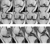
A 40-year-old man was admitted to our hospital with pain on the anteromedial side of the right knee for 15 days. The patient had no history of trauma, and physical examination revealed tenderness on the anteromedial joint line. The results of McMurray's and Apley's tests were positive on the medial side of the right knee. He complained of anterior knee pain on the hyperextension test, although he had a full range of motion. However, there was neither edema of the knee, hypotrophy of the quadriceps muscle, nor instability of the knee. Plain radiographs showed no specific abnormalities. Coronal MPD images showed a bandlike low-signal intensity with inserting into the ACL at the intercondylar notch level (Fig. 1a). Sagittal modified proton density (MPD) showed a bandlike low-signal intensity which started from the anterior horn of the medial menisucs and ran anteriorly parallel to the anteromedial bundle of the ACL (Fig. 1b). Based on MR findings, the patient was diagnosed with AIMM into the ACL. No therapeutic or diagnostic procedures, including arthroscopy, were conducted. After conservative therapy for 3 months, his pain at the right knee joint was almost relieved, and at further follow-ups, the pain completely disappeared.
DISCUSSION
Anomalies of the medial meniscus are rare. The anterior horn of the medial meniscus is the most frequent site of variation (4). Among these rare anomalies, AIMM into ACL is even more infrequent variation. In the study of Cha et al. (6) the frequency of AIMMs was 2.26% (30 MR images obtained from 1326 consecutive knee arthroscopic studies). In the normal knee, the anterior horn of the medial meniscus is attached to the tibial plateau in the area of the anterior intercondylar fossa in front of the ACL; it remains distinct from the anterior cruciate ligament. The characteristic features of AIMM into the ACL include a low-signal band that extends from the anterior horn of the medial meniscus, running parallel to the ACL up to the insertion site of the ACL or the intercondylar notch (5). Throughout the course, AIMM shows an isointense-signal intensity to the meniscus and the ACL, with a high-signal intensity gap between the ACL and the AIMM. In such anomalies, the medial meniscus has insertion into various sites: the lower part (inferior third) of the ACL, the middle part (middle third) of the ACL, and the superior part (superior third; intercondylar notch) on sagittal MR images, with the lower insertion type being more common (6).
A tear of the anterior horn of the medial meniscus or ACL and infrapatellar plicae should be considered in the differential diagnosis of AIMM. A tear in the anterior horn of the medial meniscus mimics AIMM, but AIMM can be definitively diagnosed by tracing the medial margin of the anterior horn of the medial meniscus on serial T2-weighted sagittal images. In addition, tears confined to the anterior third of the medial meniscus, particularly in its lateral part, are extremely rare, and therefore differentiation between AIMM and the tears is nearly unnecessary in clinical practice (6). Arjun et al. (5) indicated that AIMM could be mistaken for partial rupture of the ACL. However, this anomaly can be detected by tracing from the anterior horn of the medial meniscus to the ACL on thin serial sections of T2-weighted images. An infrapatellar plica is similar to AIMM, with a curvilinear appearance anterior to the ACL on sagittal T2-weighted images (7). However, the origin of the infrapatellar plica does not attach to the anterior horn of the medial meniscus but to Hoffa's fat pad or the inferior pole of the patella, which can be confirmed on MR images and arthroscopic examination. AIMM may complicate the arthroscopic evaluation of the ACL as is the case with infrapatellar plicae (7).
Cha et al. (6) hypothesized that AIMM is a variation of infrapatellar plicae because all cases showed infrapatellar plicae as thin membranes on arthroscopic examination. In addition, AIMM and infrapatellar plica have a chronological similarity in developmental stages according to recent embryological studies (8, 9).
Our case shows the typical MR findings of AIMM into the ACL. On serial sagittal and coronal images, tears of the anterior horn of the medial meniscus and ACL were excluded. Since the ACL started from the anterior aspect of the medial meniscus, our case was easily differentiated from infrapatellar plica. Generally, AIMM into the ACL does not seem to correlate with knee symptoms. Santi and Richardson (1) reported the case of a patient with bilateral anomalous insertion of the medial meniscus and the habitus of Marfan's syndrome. This patient had no other cause of the anterior medial knee pain, which disappeared after excision of the anomalous meniscal portion. In our case, after conservative treatment and regular follow-ups, the patient's pain in the knee joint was completely relieved, suggesting that the pain may not correlate with AIMM.
In conclusion, AIMM can be misdiagnosed as ACL tear, meniscal tear, or infrapatellar plica and may complicate arthroscopic findings. Familiarity with characteristic MR findings can aid in the early detection of AIMM.
 XML Download