

 PDF
PDF ePub
ePub Citation
 Print
Print


INTRODUCTION
Ectopic pancreas is defined as pancreatic tissue lacking anatomic and vascular continuity with the main body of the gland (1). Pancreatic fatty infiltration-replacement is commonly diffuse involvement, most frequently seen in elderly or obese patients, and may be associated with diseases such as chronic pancreatitis and cystic fibrosis (2). Pathogenesis of fat replacement (also termed lipomatosis, adipose atrophy, or fat infiltration) of the exocrine pancreas is not well established. In our case, peripheral steatosis was detected in ectopic pancreas, and mimicked as exogastric mass. The radiographic features of this condition have not been described, to the author's knowledge, based on a computerized search of the literature. The clinical importance of this case is to demonstrate that this finding may not be misdiagnosed as exogastric mass and highly suggesting feature for ectopic pancreas.
CASE REPORT
A 38-year-old man was admitted to our institute for evaluation of a known gastric mass. He was diagnosed it on endoscopy 2 years ago, and suffered from unintentional weight loss of 2 kg for 2 months. He had adult-onset diabetes mellitus for 3 years, which was controlled by medication. He had no history of hypertension, alcohol abuse, smoking and previous surgery. Physical examination was unremarkable. Laboratory examination revealed elevations in serum glucose 350 mg/dL (normal reference: 70-100 mg/dL), cholesterol 238 (normal reference: 238 mg/dL), and alanine transaminase (ALT) 55 IU/L (normal reference: 4-43 IU/L). Blood cell count, hemoglobin, total bilirubin, serum lipase, aspartate aminotransferase (AST), alkaline phosphatase (ALP), creatinine, and blood urea nitrogen (BUN) levels were within normal range.
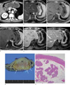
Contrast-enhanced computed tomography (CT) demonstrated 2.5 cm sized, well-defined mass with peripheral fatty infiltration on the anterior wall of the gastric lower body (Fig. 1a). The main pancreas showed normal contour and attenuation. MR images (Signa HDxt 1.5T, GE Healthcare, Milwaukee, WI) revealed a well-defined submucosal mass with smooth margins, enclosed by fat component. T2-weighted image (single shot fast spin echo sequence, TR/TE 1500/92.4 ms, flip angle 90°) demonstrated that high signal intensity margin enclose iso-signal intensity contour mass with pancreas (Fig. 1b). On T1-weighted 2D dual gradient echo (TR/TE 135/4.3 ms, flip angle 60°), inphased image (Fig. 1c) showed that hyperintense fatty infiltraion enclose iso intense lobulated mass, and opposed phase (Fig. 1d) showed signal drop on margin of fatty lesion. Non-enhanced and contrast enhanced (Omniscan, GE Healthcare) fat supressed T1-weighted images (TR/TE 4.7/2.1 ms, flip angle 12°) using the liver acquisition with volume acceleration (LAVA) sequence revealed enhancing ectopic pancreas and peripheral non-enhancing suppressed fatty component (Fig. 1e, f).
The patient underwent a gastric wedge resection including the tumor. The tumor was 5.5×3.0×3.0 cm in size, covered by gastric mucosa, arising from the muscular layer and containing pancreatic tissue with perilesional fat tissue (Fig. 1g). Histologic examination demonstrated ectopic pancreas (Fig. 1h). The postoperative course was uneventful, and he was discharged on postoperative 10 days.
DISCUSSION
Ectopic pancreas is a common developmental anomaly with a reported incidence of 0.55-14% at autopsy (1, 3, 4). They consist histologically of all pancreatic elements, including acini, islet cells, and ductal structures (2, 5, 6). Fatty replacement of pancreas may be uniform or may be unevenly distributed, and Langerhans are usually not affected by fatty replacement (4). No etiology has been established for fat replacement of the pancreas; however, several predisposing factors have been suggested: age, obesity, diabetes mellitus, chronic pancreatitis, hepatic disease, dietary deficiency, viral infection, steroid therapy, obstruction of the pancreatic duct, and fibrocystic disease (7, 8). Experimental and clinical studies have shown that fat replacement of the pancreas does result from ligation of the pancreatic duct or obstruction of the pancreatic duct by a tumor or a calculus.
We postulate that in our case, obesity and diabetes mellitus were significant etiologic factors responsible (7, 8). Hepatic disease, chronic pancreatitis and adult-onset diabetes mellitus may have been contributory (7, 8). Although the pathophysiology was not well known, uniform fatty replacement of pancreas is the most frequent CT finding in adolescent and adult patients with cystic fibrosis, and pancreatic glandular tissue is significantly reduced in size (5, 7). Fatty lesion of pancreas is also detected in cases of lipomatous pseudohypertrophy, lipoma, liposarcoma, fibrolipoma, and teratoma (4). Endoscopy may be performed to differentiate an ectopic pancreas from neoplastic lesions. Ectopic pancreas can be developed into complications such as pancreatitis, pseudocyst, insulinoma, adenoma, and malignant transformation (1, 3). These complications cause clinical symptoms such as abdominal pain, gastrointestinal bleeding, and obstruction (1, 3). Surgical resection is required for symptomatic patients with ectopic pancreas that has atypical radiographic features.
Heterotopic pancreas in the stomach may easily be misinterpreted as other gastric submucosal tumor. According to previous study (3), CT findings were interpreted correctly as heterotopic pancreas in only two (17%) cases, and the remaining 10 (83%) cases were misinterpreted as leiomyoma in four, as carcinoid tumor in three, as submucosal scirrhous carcinoma in two, and as lymphadenopathy in one (3). Although focal fatty infiltration of gastric submucosal tumor can be helpful for diagnosis of ectopic pancreas, the finding of perilesional steatosis can be detected in other tumors including lipoma, liposarcoma, angiolipoma, teratoma (9).
Prominent enhancement of the overlying mucosa, location, LD/SD ratio, growth pattern, and lesion border are statistically significant predictors in the differentiation of ectopic pancreas from GIST and leiomyoma (1). Glomus tumors appear as smooth submucosal masses with or without ulceration and may contain tiny flecks of calcification (10). These tumors frequently demonstrate strong enhancement on early-phase contrast enhanced images (10). Gastric schwannomas usually appear as discrete submucosal masses that are indistinguishable from other mesenchymal tumors (10). As they outgrow their blood supply, these lesions may undergo central necrosis and ulceration (11).
In conclusion, we report an unusual case of ectopic pancreas that appeared on radiologic images as a lobulated, submucosal mass enclosed by fat component in the gastric lower body. Although ectopic pancreas including fat component is extremely rare, in the setting of gastric submucosal mass with containing focal fat, these findings should be considered in ectopic pancreas as part of the differential diagnosis.
 XML Download