

 PDF
PDF ePub
ePub Citation
 Print
Print


Abstract
Breast hamartoma is a relatively rare pathology, composed of various amount of mammary glandular, fatty and fibrous tissue. Here, we report MR findings of two cases of hamartomas; one of them was incidentally found in her left breast during preoperative MRI in a woman with right breast cancer, and the other was presented as a large palpable mass. Both of them were confirmed by surgical excision. Breast hamartoma shows a well-defined mass with mixed signal intensity on T2-weighted image MRI and a little or focal enhancement on contrast-enhanced MRI.
Figures and Tables
Fig. 1
A 44-year-old woman with hamartoma of the left breast.
a. Axial T1-weighted spin echo MRI (TR/TE, 400/7) shows a 2 cm, oval shaped, circumscribed mass with central high signal intensity and peripheral iso signal intensity in left upper inner quadrant (arrow). b. Axial fat - suppressed fast spin echo T2-weighted MRI (TR/TE, 5000/70) shows a mass with central iso signal intensity, which was regarded as fat component and peripheral high signal intensity (arrow). c. Sagittal dynamic contrast-enhanced fat-suppressed T1-weighted image MRI shows mild peripheral nodular enhancement of the mass (arrows). d. The time-intensity curve obtained from the site of nodular enhancement during dynamic contrast enhanced MRI shows the presence of a gradual enhancement pattern. e. Second-look ultrasonogrpahy shows an oval shaped, circumscribed, hyperechoic mass in the left upper inner quadrant (arrows). f. Mediolateral mammogram shows a 2 cm, oval shaped, well-defined mass with internal fat density (arrows).
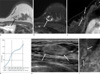
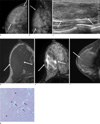
Fig. 2
A 48-year-old woman with hamartoma of the right breast.
a, b. Craniocaudal and mediolateral mammograms of her right breast show a 7cm sized, oval shaped, circumscribed, isodense mass at palpable site of the right breast (arrows). c. Ultrasonography shows an oval shaped, circumscribed, heterogeneous and isoechoic mass (arrows). d. Axial T1-weighted spin echo MRI (TR/TE, 660/8) shows an oval shape, circumscribed mass with iso signal intensity (arrows). e. Axial fat - suppressed fast spin echo T2-weighted MRI (TR/TE, 5,500/70) shows a mass with mixed signal intensity (arrows). F. Sagittal dynamic contrast-enhanced fat-suppressed T1-weighted image MRI shows no enhancement except a little peripheral enhancement (arrow). g. Photomicrograph shows several small clusters of adipocytes within a densely fibrotic stroma (adipocyte (black arrows), glandular tissue (white arrows), fibrous tissue (black arrowheads), haematoxylin and eosin stained; original magnification, ×100).
References
1. Fisher CJ, Hanby AM, Robinson L, Millis RR. Mammary hamartoma--a review of 35 cases. Histopathology. 1992. 20:99–106.
2. Charpin C, Mathoulin MP, Andrac L, et al. Reappraisal of breast hamartomas. A morphological study of 41 cases. Pathol Res Pract. 1994. 190:362–371.
3. Adler DD, Jeffries DO, Helvie MA. Sonographic features of breast hamartomas. J Ultrasound Med. 1990. 9:85–90.
4. Kievit HC, Sikkenk AC, Thelissen GR, Merchant TE. Magnetic resonance image appearance of hamartoma of the breast. Magn Reson Imaging. 1993. 11:293–298.
5. Testempassi E, Ishi C, Yamada T, Fukuda K, Tada S, Nikaido T. Case report: breast hamartoma: MR findings. Radiat Med. 1995. 13:187–189.
6. Hessler C, Schnyder P, Ozzello L. Hamartoma of the breast: diagnostic observation of 16 cases. Radiology. 1978. 126:95–98.
7. Paraskevopoulos JA, Hosking SW, Stephenson T. Breast within a breast: a review of breast hamartomas. Br J Clin Pract. 1990. 44:30–32.
8. Pui MH, Movson IJ. Fatty tissue breast lesions. Clin Imaging. 2003. 27:150–155.
9. Tse GM, Law BK, Ma TK, et al. Hamartoma of the breast: a clinicopathological review. J Clin Pathol. 2002. 55:951–954.
10. Chao TC, Chao HH, Chen MF. Sonographic features of breast hamartomas. J Ultrasound Med. 2007. 26:447–452. quiz 453.
11. Erdem G, Karakas HM, Isik B, Firat AK. Advanced MRI findings in patients with breast hamartomas. Diagn Interv Radiol. 2011. 17:33–37.
12. Kinoshita T, Yashiro N, Ihara N, Funatu H, Fukuma E, Narita M. Diffusion-weighted half-Fourier single-shot turbo spin echo imaging in breast tumors: differentiation of invasive ductal carcinoma from fibroadenoma. J Comput Assist Tomogr. 2002. 26:1042–1046.
 XML Download