

 PDF
PDF ePub
ePub Citation
 Print
Print


Abstract
Small cell carcinoma of the gallbladder is a type of neuroendocrine tumor and very rare. We report ultrasound, CT and MR findings of a small cell carcinoma of the gallbladder that was confirmed by pathology. Small cell carcinoma of the gallbladder was seen as a well-defined mass with peripheral rim enhancement in the gallbladder. In spite of the large size of the mass, direct and extensive invasion of the liver was not detected. However, there were many metastatic lymph nodes.
Figures and Tables
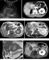 | Fig. 1A 64-year-old man with small cell carcinoma of the gallbladder.
a. Abdominal ultrasonography shows irregular shaped, heterogeneous hypoechoic solid mass (arrows) at fundus portion of the gallbladder with well-preserved lumen.
b. Contrast enhanced CT image shows the central low attenuated mass (arrows) relative to the liver with peripheral rim enhancement. There are multiple enlarged and conglomerated lymph nodes (arrowheads) with peripheral rim enhancement at hepaticoduodenal ligament.
c-f. The well-defined gallbladder mass (white arrow) at fundus portion and conglomerated lymphadenopathy (arrowheads) was shown as a large low signal intensity solid mass on T1-weighted image (c) and slightly high signal intensity relative to the liver parenchyma on T2-weighted axial (d) and coronal image (e). There was no invasion to the hepatic parenchyma. Gadolinium enhanced T1-weighted MR image (f) shows this mass (white arrow) with peripheral rim enhancement. The lumen of the gallbladder (black arrow) is relatively preserved and showed high signal intensity on T1-weighted image (c). There are multiple enlarged lymph nodes (arrowheads) at hepatoduodenal ligament and no hepatic metastasis was noted.
|
References
1. Lee MH, Park TJ, Lee HW, et al. Small cell carcinoma of the gall bladder. Two case reports. Korean J Med. 2007. 73:1022–1028.
2. Albores-Saavedra J, Cruz-Ortis H, Alcantra-Vazques A, Henson DE. Unusal types of gallbladder carcinoma. A report of 16 cases. Arch Pathol Lab Med. 1981. 105:287–293.
3. Kuwabara H, Uda H. Small cell carcinoma of the gall-bladder with intestinal metaplastic epithelium. Pathol Int. 1998. 48:303–306.
4. Moskal TL, Zhang PJ, Nava HR. Small cell carcinoma of the gallbladder. J Surg Oncol. 1999. 70:54–59.
5. Cavazzana AO, Fassina AS, Tollot M, Ninfo V. Small-cell carcinoma of gallbladder. An immunocytochemical and ultrasrtuctural study. Pathol Res Pract. 1991. 187:472–476.
6. Choi WB, Lee TY, Lee NW, et al. A case of small cell carcinoma of gallbladder. Korean J Med. 1997. 53:847–852.
7. Obuz F, Altay C, Sagol O, Astarcioglu H, Oztop I, Igci E. MDCT findings in neuroendocrine carcinoma of the gallbladder: case report. Abdom Imaging. 2007. 32:105–107.
8. Ahn JE, Byun JH, Ko MS, Park SH, Lee MG. Neuroendocrine carcinoma of the gallbladder causing hyperinsulinaemic hypoglycaemia. Clinical Radiology. 2007. 62:391–394.
 XML Download