

 PDF
PDF ePub
ePub Citation
 Print
Print


Abstract
Malignant mixed Müllerian tumors (MMMT) are rare aggressive tumors that typically arise fromthe female genital tract. This malignancy has an extremely poor prognosis due to its rapid growthand the high associated incidence of both local recurrence and distant metastases. Althoughintraperitoneal metastasis from MMMT is relatively common, no reports exist regarding theradiologic findings of intestinal metastasis from MMMT. Here, we report a case of MMMT withsecondary small bowel metastasis and the associated radiologic findings.
Figures and Tables
 | Fig. 1
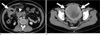
a. Contrast-enhanced axial CT image shows irregular wall thickening of the small bowel (arrow) with enlarged mesenteric and retroperitoneal lymph nodes (arrowhead).
b. Contrast-enhanced axial CT scan at the pelvic level shows a poorly enhancing mass in the uterus. The mass extends into the endometrial cavity.
|
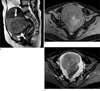 | Fig. 2(a) Sagittal T2-weighted MR image shows a large mass with slightly high signal intensity (arrows) in the uterus that extends to the uterine cervix. Pre-contrast (b) and gadolinium enhanced (c) axial fat-suppressed T1-weighted MR image shows less heterogeneous enhancement of the tumor relative to the adjacent myometrium.
|
 | Fig. 3
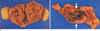
a. A photograph of the gross uterine specimen shows a poorly circumscribed infiltrative yellow, solid mass in the endomyometrium with invasion of more than one half of the myometrium.
b. The gross small bowel specimen shows an irregular mass (arrows) that is adherent to the serosal surface of the ileum.
|
References
1. Lotocki R, Rosenshein NB, Grumbine F, Dillon M, Parmley T, Woodruff JD. Mixed mullerian tumors of the uterus: clinical and pathologic correlations. Int J Gynaecol Obstet. 1982. 20:237–243.
2. Smith T, Moy L, Runowicz C. Mullerian mixed tumors: CT characteristics with clinical and pathologic observations. AJR Am J Roentgenol. 1997. 169:531–535.
3. Bharwani N, Newland A, Tunariu N, et al. MRI appearances of uterine malignant mixed mullerian tumors. AJR Am J Roentgenol. 2010. 195:1268–1275.
4. Ho SP, Ho TH. Malignant mixed mullerian tumours of the uterus: a ten-year experience. Singapore Med J. 2002. 43:452–456.
5. Montague AC, Swartz DP, Woodruff JD. Sarcoma arising in a leiomyoma of the uterus: factors influencing prognosis. Am J Obstet Gynecol. 1965. 92:421–427.
6. Zelmanowicz A, Hildesheim A, Sherman ME, et al. Evidence for a common etiology for endometrial carcinomas and malignant mixed mullerian tumors. Gynecol Oncol. 1998. 69:253–257.
7. Lim BJ, Kim JW, Yang WI, Cho NH. Malignant mixed mullerian tumor of fallopian tube with multiple distinct heterologous components. Int J Gynecol Cancer. 2004. 14:690–693.
8. Sahdev A, Sohaib SA, Jacobs I, Shepherd JH, Oram DH, Reznek RH. MR imaging of uterine sarcomas. AJR Am J Roentgenol. 2001. 177:1307–1311.
9. Teo SY, Babagbemi KT, Peters HE, Mortele KJ. Primary malignant mixed mullerian tumor of the uterus: findings on sonography, CT, and gadolinium-enhanced MRI. AJR Am J Roentgenol. 2008. 191:278–283.
10. Ko EY, Ha HK, Kim AY, et al. CT differentiation of mucinous and nonmucinous colorectal carcinoma. AJR Am J Roentgenol. 2007. 188:785–791.
11. Callister M, Ramondetta LM, Jhingran A, et al. Malignant mixed Müllerian tumors of the uterus: analysis of patterns of failure, prognostic factors, and treatment outcome. Int J Radiat Oncol Biol Phys. 2004. 58:786–796.
 XML Download