

 PDF
PDF ePub
ePub Citation
 Print
Print


Abstract
Anoperineal tuberculosis is a rare extrapulmonary form of the disease and may present as abscess. We report a case of anoperineal tuberculous abscess, which showed low signal intensity on T1-weighted images, high signal intensity on T2-weighted images and diffusion restriction on diffusion weighted images.
Figures and Tables
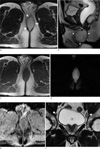
Fig. 1
(a) Axial and (b) sagittal T2-weighted images show well-circumscribed oval shaped hyperintense mass (arrow heads) in the perineum with peripheral hypointense rim and internal septations. The mass extends into intersphincteric space (arrow) superiorly.
c. On axial T1-weighted images, the mass shows low signal intensity.
d. Diffusion weighed image at b=800 sec/mm2 shows prominent internal hyperintensity in the mass.
e. ADC map reveals diffusion restriction in the mass.
f. The oval shaped and enlarged lymph nodes (arrow heads) were seen along the bilateral pelvic wall.
References
1. Raviglione MC, Snider DE Jr, Kochi A. Global epidemiology of tuberculosis. Morbidity and mortality of a worldwide epidemic. JAMA. 1995. 273:220–226.
2. Harland RW, Varkey B. Anal tuberculosis: report of two cases and literature review. Am J Gastroenterol. 1992. 87:1488–1491.
3. Burrill J, Williams CJ, Bain G, Conder G, Hine AL, Misra RR. Tuberculosis: a radiologic review. Radiographics. 2007. 27:1255–1273.
4. Candela F, Serrano P, Arriero JM, Teruel A, Reyes D, Calpena R. Perianal disease of tuberculous origin: report of a case and review of the literature. Dis Colon Rectum. 1999. 42:110–112.
5. Yaghoobi R, Khazanee A, Bagherani N, Tajalli M. Gastrointestinal tuberculosis with anal and perianal involvement misdiagnosed as Crohn's disease for 15 years. Acta Derm Venereol. 2011. 91:348–349.
6. Jinkins JR, Gupta R, Chang KH, Rodriguez-Carbajal J. MR imaging of central nervous system tuberculosis. Radiol Clin North Am. 1995. 33:771–786.
7. Murata Y, Yamada I, Sumiya Y, Shichijo Y, Suzuki Y. Abdominal macronodular tuberculomas: MR findings. J Comput Assist Tomogr. 1996. 20:643–646.
8. Morita S, Higuchi M, Takahata T, et al. Magnetic resonance imaging for multiple macronodular localized splenic tuberculosis. Clin Imaging. 2007. 31:134–136.
9. Luthra G, Parihar A, Nath K, et al. Comparative evaluation of fungal, tubercular, and pyogenic brain abscesses with conventional and diffusion MR imaging and proton MR spectroscopy. AJNR Am J Neuroradiol. 2007. 28:1332–1338.
10. Tappouni RF, Sarwani NI, Tice JG, Chamarthi S. Imaging of unusual perineal masses. AJR Am J Roentgenol. 2011. 196:W412–W420.
 XML Download