

 PDF
PDF ePub
ePub Citation
 Print
Print


Abstract
Figures and Tables
Fig. 1
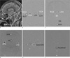
Fig. 2
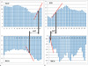
Fig. 3
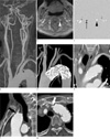
Fig. 4
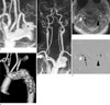
Table 2
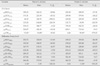
Note.- TEVAR, thoracic endovascular aortic repair; %Δ = (FV after TEVAR - FV before TEVAR) / FV before TEVAR × 100 (%); C1-2, between first and second cervical vertebral level; MBA, midbasilar artery level; RICA, right internal carotid artery; LICA, left internal carotid artery; RVA, right vertebral artery; LVA, left vertebral artery; TCBF, total cerebral blood flow; IJV, internal jugular vein; CSF, cerebrospinal fluid; BA, basilar artery; FVSS+SSSax or FVSS+SSScor, sum of FVs at straight sinus and superior sagittal sinus measured at axial or coronal plane; FVTS, sum of FVs measured at right and left transverse sinus; FVIJV, sum of FVs measured at right and left internal jugular vein; FVCSFC1-2 or FVCSFaqueduct, CSF flow measured at C1-2 or aqueduct; *, minus means caudocephalad direction of averaged FVCSFC1-2 and FVCSFaqueduct.
Table 3
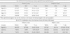
Note.- TEVAR, thoracic endovascular aortic repair; % Δ = (AVTT after TEVAR - AVTT before TEVAR) / AVTT before TEVAR × 100 (%); ICA, internal carotid artery; VA, vertebral artery; IJV, internal jugular vein; BA, basilar artery; SS, straight sinus; SSS, superior sagittal sinus.**0, Total trigger delay at this plane was 532 msec in patient 1.
 XML Download