

 PDF
PDF ePub
ePub Citation
 Print
Print


Introduction
Lung cancer is one of the most common malignancies and the leading cause of cancer death. The welle-stablished therapeutic methods for a primary lung malignancy or secondary pulmonary metastatic nodules are surgery, chemotherapy, and radiation therapy. Unfortunately, patients with a lung malignancy or pulmonary metastatic nodules are often poor surgical or chemotherapy candidates due to chronic obstructive pulmonary disease, co-morbidity, or cardiac disease (1-5).
Most investigators have been used CT as a follow up modality after RFA (4-9). Steinke et al. (7) reported the complete absence of contrast enhancement in the ablated zones on the follow-up CT scans obtained immediately after treatment in the complete ablation group, whereas the ablated zones in the partial ablation group showed various degrees of enhancement.
MRI has been explored as an alternative imaging method for lung cancer, with the potential to provide morphological information, while being completely free from radiation hazards. To our knowledge, there were a few of the MRI findings of the ablated zones after RFA in patients with unresectable lung malignancies (10, 11). Okuma et al. (10) reported that the apparent diffusion coefficient (ADC) on DWI without local progression was significantly higher than with local progression after RFA, suggesting that the ADC can predict the response to RFA for lung tumors. However, there is no report for local progression of ablated lesion using multiphase contrast-enhanced MRI (CE-MRI).
In this study, we examined which phase was important for predicting local recurrence of an ablated zone after RFA treatment in patients with an unresectable lung malignancy on multiphase CE-MRI.
Materials and Methods
Patients
The MRI assessment of the ablated zone after RFA was retrospectively investigated with the approval of the institutional ethics committee at our institution. Sixteen patients with a primary lung malignancy and four patients with a single metastatic nodule from extrapulmonary malignancies were referred for CT-guided RFA. There were sixteen men and four women, ranging in age from 37 to 84 years (mean age, 61.5 years). All patients were diagnosed non-small cell lung cancer using percutaneous needle lung biopsy or bronchoscopic biopsy and included as follows; (a) medical inoperability (stage I - II) due to medical contraindications or strong refusal of surgery (n=5); (b) the presence of a single metastatic nodule originating from extrapulmonary malignancy (n=4); (c) the patients want to reduce the volume of the primary tumor in patients with stage III-IV (n=11).
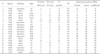
The tumors ranged in diameter from 1 to 5 cm (mean, 3.9±1.1 cm). The tumor cell types were squamous cell carcinomas (n=10), adenocarcinomas (n=6), and metastases (n=4; colon, breast, uterus). Five patients (25%) had stage Ia or Ib lung cancer, eleven (55%) had an unresectable stage III or IV cancer, and four (20%) had metastases from an extrapulmonary malignancy (Table 1).
RFA Preparation and Procedure
RFA was performed on in-patients after 6 hrs fasting. The coagulation parameters were always checked before the procedure. All patients underwent a chest CT examination (Somatom Plus 4, Siemens, Erlargen, Germany) at most one week before RFA. RFA was always performed by one radiologist with CT guidance. The patients' vital signs were monitored continuously throughout the procedure. In all the patients, RF therapy was performed with the analgesia achieved by the intravenous administration of 50-100 µg of fentanyl citrate (Myungmoon, Kyungki, Korea). RFA was carried out using a 17-gauge, single internally cooled RF electrode (Radionics, Burlington, Mass, USA). Once proper positioning was confirmed, the electrode was attached to a 500-KHz, monopolar RF generator (CC-1; Radionics, Burlington, MA, USA). At the end of each treatment, the perfusion was stopped and the maximum temperature was recorded. If the temperature exceeded 60℃ during treatment, the electrode was withdrawn in increments of 1 cm up to the length of the active-tip, while the intratumoral temperature was measured simultaneously. An additional treatment was performed at the same position if the maximum intratumoral temperature recorded after the RFA treatment did not exceed 60℃. After treatment, the electrode was withdrawn without cauterizing the probe tract (2, 9).
MR Sequence and Analysis of the MR Findings
The chest MRI images were obtained within 24 hrs after RFA treatment to assess the immediate results. The post-RFA MRI (Magnetom Symphony, Siemens, Erlargen, Germany) was evaluated by two radiologists, who are experienced in chest CT scans and MRI and were aware of the findings of the CT scans performed before RFA.
In all cases, MRI was performed on a 1.5-T superconducting imager using phased array multicoils. The lungs were imaged using the following sequence: contrast-enhanced T1-weighted image (volumetric interpolated breath-hold examination, VIBE). CE-MRI using VIBE was performed after administering gadopentate dimeglumine (Gd-BOPTA; MultiHance, Bracco SpA, Milan, Italy) at a dose of 0.1 mmol/kg and a rate of 3 cc/sec using an MR-compatible power injector (Spectris®; Medrad, Pittsburgh, USA). VIBE was performed in the pre-contrast, arterial (20-35 sec), venous (45-60 sec), and equilibrium phases (3-5 min) using the following parameters: 3.4/1.5 msec; flip angle, 12°; bandwidth, 490 Hz/pixel; matrix, 256 (readout direction) × 120 (phase) × 64-72 (partition); effective slice thickness, 2.3 mm; and field of view, 30-35 cm (12, 13). Image reconstruction was carried out using the source images taken with a slice thickness of 3 mm on an MRI workstation.
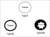
Image analysis was performed independently by two radiologists, who analyzed the CT images obtained before RFA and the MRI findings after RFA on the same patient using a picture archiving and communication system (PACS, m-view™; Marotech, Korea). The enhancement pattern on CE-MRI was also analyzed. The ablated zone was divided into the central zone (central ablated zone except for the ablated margin) and the ablated margin (peripheral ablated zone within 10 mm of the outermost margin of the ablated zone). These enhancement patterns were classified into the following three types: Type I, the ablated zone showed absolutely no enhancement; Type II, only enhancement of the ablated margin was observed; and Type III, heterogeneous enhancement of the ablated zone (Fig. 1).
The tumor sizes before and after treatment were assessed in the axial plane using the maximum diameters on the pre procedural CT image taken in the axial plane and the follow-up contrast-enhanced CT, respectively.
Therapeutic Response Criteria
For the therapeutic response, complete ablation was defined as no enhancement (type I) of the ablated lesion at CE-MRI obtained within 24 hours after RFA. Also, there was decrease or no change of the size, or no enhancement of the treated lesion at last follow-up contrast-enhanced CT. Unsuccessfully treated was defined as enhanced lesion at CE-MRI (type II or III) obtained within 24 hours after RFA. Additionally, there was an increase in size of ablated lesion at last follow-up contrast-enhanced CT.
The tumor size was assessed in the transverse plane with the largest diameter and the greatest perpendicular measurement recorded on the post-RFA MRI and follow-up contrast CT. The CE-MRI obtained within 24 hours after RFA compared with baseline contrast-enhanced CT scans that obtained before the procedure, and technical success assessed. Technical success, primary effectiveness rate, and local tumor progression were defined according to the guidelines recommended by Goldberg et al. [14] Technical success was defined when tumor was treated according to protocol and completely tumor coverage can be assessed immediately after the procedure on multiphase CE-MRI. Also, primary effectiveness rate is defined as the percentage of tumors that were successfully eradicated following the initial procedure. We evaluated primary effectiveness rate and local tumor progression as performed using a contrast-enhanced CT at 1, 3 months and every 6 months after RFA.
Statistical Analysis
A κ coefficient was calculated to evaluate the degree of agreement between the two chest radiologists in evaluating lung cancer on CT and ablated lesion after RFA (less than 0.20, poor agreement; 0.21-0.40, fair agreement; 0.41-0.60, moderate agreement; 0.61-0.80, good agreement; and 0.81-1.00, very good agreement). The Spearman correlation coefficient was also used to characterize the MR features (type of ablated lesion) and local tumor progression of the ablated zone on the follow up CT. Also, we evaluated diagnostic performance of CE-MRI (arterial, venous, or equilibrium phase) for local recurrence using chi-square test when predicted local recurrence of CE-MRI was defined type III pattern. All analyses were performed using SPSS 9.0 computer software (SPSS Inc., Chicago, Ill., U.S.A). The values are expressed as a mean ± SD. For all statistical analyses, p values < 0.05 were considered significant.
Results
All patients (n=20) treated by RFA were technical successes (100%). On follow-up CT, primary effectiveness rate were shown in 9 patients (45%) and 11 patients (55%) were shown to have local progression. The overall survival time for the patients with lung malignancy in this study were 13.1±6.8 months. Of the 20 patients, nine (45%) showed a gradual decrease or no change in the diameter of the ablated zone on contrast-enhanced CT until the last follow-up (mean, 17.1±5.3 months); mean size, 2.5±1.0 cm to 2.4±1.4 cm. Eleven patients (55%) showed a gradual increase in the diameter of the ablated zone on contrast-enhanced CT until the last follow-up (mean, 9.7±6.2 months); mean size, 4.7±1.1 cm to 6.8±2.0 cm (Table 1).
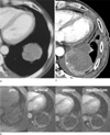
In the arterial phase on CE-MRI after RFA, the enhancement of the ablated zone was categorized as types I (n=1, 5%), II (n=13, 65%), and III (n=6, 30%). In the venous phase, the enhancement of the ablated zone was categorized as types I (n=1, 5%), II (n=8, 40%), and III (n=11, 55%). In the equilibrium phase, the enhancement of the ablated zone was categorized as types II (n=7, 35%) and III (n=13, 65%) (Figs. 2, 3). Interobserver agreement of the enhancement patterns of the lung cancers on the contrast-enhanced CT [κ (95% CI) values of 0.825 (0.650-0.934)] and the enhancement of the ablated zone on CE-MRI after RFA [κ (95% CI) values of 0.715 (0.530-0.872)] was assessed using the kappa test, which indicated a good and moderate inter-observer correlation, respectively.
Of the 20 patients, six (30%) showed a complete ablation and fourteen (70%) had unsuccessfully treated. In those with a complete ablation, the enhancement pattern was types I (11.1%) or II (88.9%) regardless of the enhancement time. In the case of unsuccessfully treated, type II (57.1%) or III (42.9%) pattern observed in the arterial phase. However, types II (21.4%) and III (78.6%) observed in the venous, and types II (7.1%) and III (92.9%) observed equilibrium phases. Sensitivity, specificity, PPV, NPV, and accuracy of local recurrence of ablated lesion on arterial phases of CE-MRI were 100%, 42.9%, 42.9%, 100%, and 60%, respectively. Sensitivity, specificity, PPV, NPV, and accuracy of local recurrence of ablated lesion on venous phases of CE-MRI were 100%, 66.7%, 78.6%, 100%, and 85%, respectively. Sensitivity, specificity, PPV, NPV, and accuracy of local recurrence of ablated lesion on equilibrium phases of CE-MRI were 100%, 85.7%, 92.9%, 100%, and 95%, respectively. The Spearman's ranked test showed a correlation between local progression and the enhancement pattern of the ablated zone in the arterial, venous, and equilibrium phases, respectively (r = 0.7, r = 0.72, r = 0.8, p < .05). Base on these results, we suggested that the equilibrium phase is important in predicting the recurrence after RFA in patients with a lung malignancy than the arterial and venous phase on the CE-MRI.
Discussion
CT is still the main imaging technique for evaluating the treatment response of a lung malignancy after RFA. As another modality for a follow-up post-RFA image, some authors (15-19) reported that post RFA PET showed virtually no FDG activity in the previous positive areas. In addition, they stated that MRI or dynamic MRI revealed reduced or no enhancement of the ablated zone. These findings corresponded to the early changes in coagulative necrosis on the histopathologic examination in the acute phase after RFA. In this study, although the enhancement pattern of ablated zone in all phase correlated therapeutic response, equilibrium phase CE-MRI provided more information whether the ablated zone would be recurrent lung cancer or not. When type III was observed in the equilibrium phase on multiphase CE-MRI, all patients have been recurred lung malignancy on follow-up CT.
Multiphase CE-MRI has two advantages in evaluating the ablated zone after a RFA treatment for a lung malignancy over contrast-enhanced CT. First, CE-MRI might be superior to contrast-enhanced CT for evaluating the ablated margin and ablated zone because MRI shows superior tissue contrast to CT. The second advantage is that CE-MRI may be useful for evaluating patients who cannot tolerate iodinated contrast agents (10, 11). If contrast-enhanced CT cannot be performed after RFA, there might be some hesitation in using RFA to treat lung malignancies because of the consequent inability to evaluate the therapeutic response. Although PET or PET/CT can provide more detailed information on the ablated zone, this modality is more expensive than CT or MRI. As a result, CE-MRI is recommended as a follow-up image modality for evaluating the ablated zone after RFA in patients with renal failure or an allergy to the CT contrast agent.
An evaluation of the ablated margin and discrimination between the hyperemic zone and remnant tumor using CT, MRI or PET/CT is very important because it is closely associated with the degree of ablation after RFA (3). Hence, attempts should be made to identify any remnant tumor with an ablated margin using imaging modalities. In this study, the important factor for predicting of therapeutic response after RFA was found to be the enhancement patterns on CE-MRI. If the size of the ablated zone was larger than the original tumor, the enhanced region outside of the enveloped low signal rim must be a hyperemic region. Enhancement of the hyperemic region was categorized as type II in this study. In the venous phase on CE-MRI, patients who showed a type II pattern became complete ablation (62.5%) or unsuccessful treated (37.5%) at the last follow-up CT. Of these patients, 25% patients changed the enhanced pattern to type III on the equilibrium phase CE-MRI, and the tumor size increased on the last follow-up CT. As a result, it is possible to include the remnant tumor within the ablated zone if the enhancement pattern changed from type II to type III on multiphase CE-MRI.
This study had a limitation. There was no comparison of the enhancement pattern of the ablated zone on MRI before and after RFA because pre-RFA MRI was not performed. Therefore, such a comparison will be needed to accurately evaluate the ablated margin and access the therapeutic efficacy after RFA in the future.
In conclusion, CE-MRI is a useful imaging modality for evaluating the ablated zone after RFA. In addition, the equilibrium phase can play a more important role in predicting the recurrence after RFA in patients with a lung malignancy than the arterial or venous phase on the CE-MRI.

 XML Download