

 PDF
PDF ePub
ePub Citation
 Print
Print


Abstract
Metastases to the orbit usually affect the intraorbital fat and bone than the extraocular muscles. Metastasis to the extracoular muscles commonly occurs unilaterally, and diffuse enlargement of the bilateral extraocular muscles due to metastasis is extremely rare. In this report, we will describe a case of diffuse metastasis to the bilateral extraocular muscles from nasal rhabdomyosarcoma masquerading as thyroid associated orbitopathy. We will also discuss about the MR imaging findings helpful for differential diagnosis from thyroid associated orbitopathy.
Figures and Tables
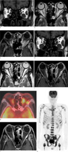
Fig. 1
(a-c) Initial orbit MR. Coronal T1-weighted (a), axial T2-weighted (b) and contrast-enhanced axial T1-weighted (c) images show diffuse enlargement and mild enhancement of the extraocular muscles except the left superior and the right lateral rectus muscles with sparing of the tendinous parts. The fat plane is clearly defined between the extraocular muscles and the orbital fat. Note there is abrupt curving and nodularity of the dilated muscle belly at the tendinous portion of the left medial rectus muscle (arrow). (d-f) Follow-up orbit MR 25 days after steroid pulse therapy. In addition to marked enlargement of the extraocular muscles on coronal T1-wieghted image (d), focal nodularity of the involvement muscles are more clearly demonstrated on axial T2-weighted (e) and contrast-enhanced axial T1-weighted (f) images. (g-h) 18F-FDG PET/CT performed on the same day with the 2nd follow-up orbit MRI. An axial 18F-FDG PET/CT image (g) shows intense hypermetabolism of the thickened bilateral medial and left lateral rectus muscles (SUVmax, 6.0 - 7.4 g/mL). The coronal maximum intensity projection image (h) shows multifocal hypermetabolic areas involving the axial and appendicular skeleton from disseminated bone metastasis. Contrast-enhanced axial T2-weighted image (i) after additional chemotherapy shows remarkable regression of the metastatic tumor involving the bilateral extraocular muscles.
References
1. Lacey B, Chang W, Rootman J. Nonthyroid causes of extraocular muscle disease. Surv Ophthalmol. 1999. 44:187–213.
2. Amato MM, Esmaeli B, Shore JW. Orbital rhabdomyosarcoma metastatic to the contralateral orbit: a case report. Ophthalmology. 2002. 109:753–756.
3. Hatton MP, Green L, Boulos PR, Rubin PA. Rhabdomyosarcoma metastases to all extraocular muscles. Ophthal Plast Reconstr Surg. 2008. 24:336–338.
4. Gupta P, Singh U, Singh SK, Kapoor R, Gupta V, Das A. Bilateral symmetrical metastasis to all extraocular muscles from distant rhabdomyosarcoma. Orbit. 2010. 29:146–148.
5. Shields JA, Bakewell B, Augsburger JJ, Flanagan JC. Classification and incidence of space-occupying lesions of the orbit. A survey of 645 biopsies. Arch Ophthalmol. 1984. 102:1606–1611.
6. Lell M, Schulz-Wendtland R, Hafner A, Magener A, Bautz WA, Tomandl BF. Bilateral orbital tumour as the presentation of mammographically occult breast cancer. Neuroradiology. 2004. 46:682–685.
7. Capone A, Slamovits TL. Discrete metastasis of solid tumors to extraocular muscles. Arch Ophthalmol. 1990. 108:237–243.
8. Jones IS, Reese AB, Krout J. Orbital rhabdomyosarcoma: an analysis of sixty-two cases. Trans Am Ophthalmol Soc. 1965. 63:223–255.
9. Mafee MF. Som PM, editor. Orbit: embryology, anatomy, and pathology. Head and neck imaging. 2003. 4th ed. St. Louis: Mosby;591–595.
10. Burch HB, Wartofsky L. Graves ophthalmopathy: current concepts regarding pathogenesis and management. Endocr Rev. 1993. 14:747–793.
 XML Download