

 PDF
PDF ePub
ePub Citation
 Print
Print


Abstract
Purpose
To describe normal anatomy and compare the differences of external genital organs and urethra on MR imaging in pre- and postmenopausal women.
Materials and Methods
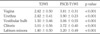
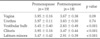
A total of 19 pre- and 18 postmenopausal healthy women underwent pelvis MR imaging at 1.5 T. Two radiologists retrospectively scored and compared the image quality of female external genitalia and urethra on axial T2-weighted images (T2WI) and axial fat-suppressed contrast-enhanced T1-weighted images (FSCE-T1WI) by using Wilcoxon signed ranks test. The radiologists compared the wall thickness or size of external genital organs and urethra on FSCE-T1WI between two groups by using Student t test.
Results
Image quality was better with FSCE-T1WI than with T2WI in all subjects (p < 0.05). The vestibular bulb, clitoris and labium minor were more clearly visualized on FSCE-T1WI in premenopausal subjects rather than in postmenopausal subjects (p < 0.05). The urethra had a target-like appearance with three layers in premenopausal and postmenopausal subjects. Postmenopausal subjects were observed to have significantly smaller vaginal wall thickness, urethral wall thickness and vestibular bulb width than premenopausal subjects (p < 0.05).
Figures and Tables
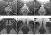 | Fig. 1Fat-suppressed contrast-enhanced axial T1 weighted (a-c) and T2-weighted (d-f) MR images of genitalia in a premenopausal subject. All genital structures were delineated more clearly on fat-suppressed contrast-enhanced axial T1-weighted image. Cc, clitoral crus. Ch, clitoral hood. Cg, clitoral glans. Lm, labia minora. U, urethra. V, Vagina. Vb, Vestibular bulb.
|
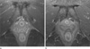 | Fig. 2Fat-suppressed contrast-enhanced T1 weighted images at pubic symphysis level in premenopausal (a) and postmenopausal (b) subjects. The separate three layers of vaginal wall are well visualized in a premenopausal subject but not clearly defined in a postmenopausal subject.
|
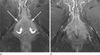 | Fig. 3Fat-suppressed contrast-enhanced T1 weighted images at ischial tuberosity level in premenopausal (a) and postmenopausal (b) subjects. The clitoral crura (straight arrow) are well delineated as a wishbone-shaped structure surrounding the urethra and vagina. The vestibular bulb (curved arrow) is paramedian in location and lying just posterior to the clitoral crura. The vestibular bulb and clitoral crura are more clearly visualized in a premenopausal subject rather than in a postmenopausal subject.
|
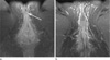 | Fig. 4Fat-suppressed contrast-enhanced T1 weighted images at vaginal introitus level in premenopausal (a) and postmenopausal (b) subjects. The clitoral glans (arrow) and labium minora are more clearly visualized in a premenopausal subject rather than in a postmenopausal subject. The anterior borders of labium minora form the clitoral hood which covers the clitoral glans.
|

References
1. Siegelman ES, Outwater EK, Banner MP, Ramchandani P, Anderson TL, Schnall MD. High-resolution MR imaging of the vagina. Radiographics. 1997. 17:1183–1203.
2. Suh DD, Yang CC, Cao Y, Garland PA, Maravilla KR. Magnetic resonance imaging anatomy of the female genitalia in premenopausal and postmenopausal women. J Urol. 2003. 170:138–144.
3. Strohbehn K, Quint LE, Prince MR, Wojno KJ, Delancey JO. Magnetic resonance imaging anatomy of the female urethra: a direct histologic comparison. Obstet Gynecol. 1996. 88:750–756.
4. Hricak H, Chang YC, Thurnher S. Vagina: evaluation with MR imaging. Part I. Normal anatomy and congenital anomalies. Radiology. 1988. 169:169–174.
5. Deliganis AV, Maravilla KR, Heiman JR, Carter WO, Garland PA, Peterson BT. Female genitalia: dynamic MR imaging with use of MS-325: initial experiences evaluating female sexual response. Radiology. 2002. 225:791–799.
6. Maravilla KR, Cao Y, Heiman JR, et al. Noncontrast dynamic magnetic resonance imaging for quantitative assessment of female sexual arousal. J Urol. 2005. 173:162–166.
7. O'Connell HE, DeLancey JO. Clitoral anatomy in nulliparous, healthy, premenopausal volunteers using unenhanced magnetic resonance imaing. J Urol. 2005. 173:2010–2063.
8. Chang SD. Imaging of the vagina and vulva. Radiol Clin North Am. 2002. 40:637–658.
9. Tan IL, Stoker J, Zwamborn AW, Entius KA, Calame JJ, Laméris JS. Female pelvic floor: endovaginal MR imaging of normal anatomy. Radiology. 1998. 206:777–783.
10. Hendren WH. Pediatric rectal and perineal problems. Pediatr Clin North Am. 1998. 45:1353–1372.
11. O'Connell HE, Hutson JM, Anderson CR, Plenter RJ. Anatomical relationship between urethra and clitoris. J Urol. 1998. 159:1892–1897.
12. Schaffer J, Fantl JA. Urogenital effects of the menopause. Baillieres Clin Obstet Gynaecol. 1996. 10:401–417.
13. Brincat M, Kabalan S, Studd JW, Moniz CF, de Trafford J, Montgomery J. A study of the decrease of skin collagen content, skin thickness, and bone mass in the post menopausal woman. Obstet Gynecol. 1987. 70:840–845.
14. Forsberg JG. A morphologist's approach to the vagina - age-related changes and estrogen sensitivity. Maturitas. 1995. 22:Suppl. S7–S15.
15. Basaran M, Kosif R, Bayar U, Civelel B. Characteristics of external genitalia in pre-and post menopausal women. Climacteric. 2008. 11:416–421.
16. Losif CS. Effects of protracted administration of estriol on the lower genitor urinary tract in postmenopausal women. Arch Gynecol Obstet. 1992. 251:115–120.
 XML Download