

 PDF
PDF ePub
ePub Citation
 Print
Print


Abstract
Granulocytic sarcoma is a manifestation of myelogenous leukemia, which means a solid mass consisting of primitive precursors of the granulocytic series of white blood cells. We present CT and MR imaging findings of bilateral sino-orbital granulocytic sarcoma in a 22-month-old boy. The mass involved bilateral orbital fossa which resulted in bilateral proptosis. Moreover, the mass extended to the almost skull base including paranasal sinuses, maxilla, temporal bone, zygomatic bone, sphenoid bone, ethmoid, and palatine bone. The adjacent dura was continuously thickened and the lower half of cavernous sinus was also involved. The patient was diagnosed as AML (M5) with t(8,21) translocation through a chromosome study from the bone marrow.
Figures and Tables
Fig. 1
Orbital CT in a 22-month-old boy with bilateral proptosis.
(Left) Non-enhanced coronal reformatted CT in a soft tissue window setting demonstrated hyperdense masses in bilateral inferolateral walls of orbits, PNS, maxillae and temporal regions.
(Right) Coronal reformatted CT in a bone window setting presented expansion of medullary cavity of frontal bone, maxilla and zygoma and no destruction of bony cortex. Most of skull base including temporal, sphenoid, palatine and ethmoid bones was also involved (not shown).
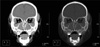
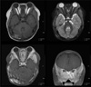
Fig. 2
Contrast-enhanced orbital MR in the same patient.
T1-weighted (Left upper) and T2-weighted (Right upper) axial images at the level of optic nerve showed iso-intense masses surrounding the bilateral lesser wings of sphenoid bone. On contrast-enhanced T1-weighted axial image (Left lower), the masses were homogeneously and strongly enhanced. Coronal image (Right lower) at the maxillary infundibular level presented homogeneous and strong enhancement in the bilateral peri-orbital soft tissue, temporal bone, maxilla, zygoma and surrounding soft tissue.
References
1. Pui MH, Fletcher BD, Langston JW. Granulocytic sarcoma in childhood leukemia: imaging features. Radiology. 1994. 190(3):698–702.
2. Pui CH. Childhood leukaemias. 1999. Cambridge: cambridge university press;288–312. 443–481.
3. Binder C, Tiemann M, Haase D, Humpe A, Kneba M. Isolated meningeal chloroma (granulocytic sarcoma) - a case report and review of the literature. Ann Hematol. 2000. 79(8):459–462.
4. Bulas RB, Laine FJ, Das Narla L. Bilateral orbital granulocytic sarcoma (chloroma) preceding the blast phase of acute myelogenous leukemia: CT findings. Pediatr Radiol. 1995. 25(6):488–489.
5. Guermazi A, Feger C, Rousselot P, et al. Granulocytic sarcoma (chloroma): imaging findings in adults and children. AJR Am J Roentgenol. 2002. 178(2):319–325.
6. Chung EM, Murphey MD, Specht CS, Cube R, Smirniotopoulos JG. From the Archives of the AFIP. Pediatric orbit tumors and tumorlike lesions: osseous lesions of the orbit. Radiographics. 2008. 28(4):1193–1214.
7. Jakobiec FA. Granulocytic sarcoma. AJNR Am J Neuroradiol. 1991. 12(2):263–264.
8. Stein-Wexler R, Wootton-Gorges SL, West DC. Orbital granulocytic sarcoma: an unusual presentation of acute myelocytic leukemia. Pediatr Radiol. 2003. 33(2):136–139.
9. Uyesugi WY, Watabe J, Petermann G. Orbital and facial granulocytic sarcoma (chloroma): a case report. Pediatr Radiol. 2000. 30(4):276–278.
 XML Download